Thoracic cavity
Question 1. Define the thoracic cavity and give its boundaries and contents.
Answer:
Thoracic Cavity:
It is a cavity of the thorax. It is enclosed in an elastic osseocartilaginous framework which helps in both increasing and decreasing the volume of the thoracic cavity.
Thoracic Cavity Boundaries:
- Anterior: Sternum.
- Lateral: 12 ribs with their costal cartilages and intercostal spaces containing intercostal muscles, membranes, nerves, and vessels.
- Posterior: Bodies of 12 thoracic vertebrae and the intervening intervertebral discs.
Read And Learn More: Anatomy Question And Answers
Thoracic Cavity Contents: The major contents are
- Lungs, chief organs of respiration
- Heart, the chief organ of circulation
- Trachea and esophagus
- Major blood vessels associated with the heart
Question 2. Write a short note on the thoracic inlet.
Answer:
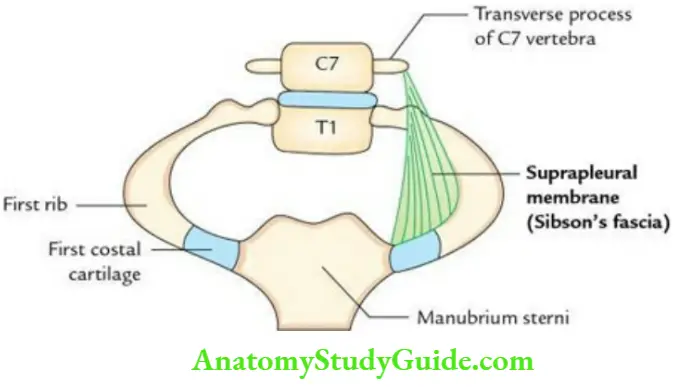
Thoracic Inlet:
It is a superior aperture of the thoracic cavity. It is reniform in shape and measures 10 cm in the transverse plane and 5 cm in the anteroposterior plane. The inlet slopes downwards and anteriorly at an angle of 45°. It is partially closed on each side by a subpleural membrane (also called Sibson’s fascia).
Thoracic Inlet Boundaries:
- Posterior: Body of first thoracic vertebra.
- On each side: First rib and its costal cartilage.
- Anterior: Upper border of manubrium sterni.
Question 3. Enumerate the structures passing through the thoracic inlet.
Answer:
The major structures are:
1. Two tubes: Trachea and esophagus
2. Two sets of arteries:
- Branches of the arch of the aorta viz. brachiocephalic trunk left common carotid and left subclavian
- Right and left internal thoracic arteries
3. Four sets of neural structures:
- Right and left vagus nerves
- Right and left phrenic nerves
- Right and left sympathetic trunks
- Right and left first thoracic nerves (ventral rami)
Note: Apices of the lungs covered by the cervical pleura also project upward through the inlet into the root of the neck.
Question 4. Write a short note on the Suprapleural membrane (Sibson’s fascia).
Answer:
It is a tough triangular membrane, which on either side partly separates the thoracic cavity from the neck.
Suprapleural membrane Features:
- Its apex is attached to the tip of the transverse process of the seventh cervical (C7) vertebra.
- Its base is attached to the inner border of the first rib and its cartilage.
- Its inferior surface is fused with the cervical pleura.
- Its superior surface is related to subclavian vessels.
Note: Morphologically, Sibson’s fascia represents the degenerated tendon of the scalenus minimus (pleuralis) muscle.
Applied anatomy:
- It protects the apex of the lung from injury.
- It prevents the puffing of the root of neck during respiration.
Question 5. Describe the thoracic outlet in brief.
Answer:
It is the inferior aperture of the thoracic cavity and is closed completely by the thoracoabdominal diaphragm (or diaphragm).
Thoracic outlet Boundaries:
- Anterior: Xiphoid process.
- Anterolaterally: Costal margin formed by the union of 7th to 9th costal cartilages.
- Posterolaterally: Tips of 11th and 12th ribs.
- Posterior: Body of T12 vertebra.
Note: The structures forming the boundaries of the thoracic outlet form an osteocartilaginous rim for the origin of the thoracoabdominal diaphragm.
The structures freely pass to and fro between thoracic and abdominal cavities through openings in the thoracoabdominal diaphragm.
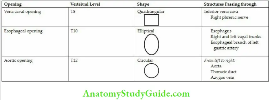
Question 6. Enumerate the major (large) openings in the diaphragm and structures passing through them in tabular form.
Answer:
There are 3 major (large) openings in the diaphragm. The details of these openings and structures passing through them are given in Table.
Major Openings of the Diaphragm and Structures Passing through Them:
Question 7. Write a short note on the cervical rib
Answer:
It is an extra rib that develops from a costal element of the transverse process of the C7 vertebra. It is present in about 0.2–0.5% of the cases. Its posterior end is attached to the transverse process of the C7 vertebra, and its distal extremity is free or attached to the first rib.
Cervical rib Applied anatomy:
The cervical rib reduces the size of the scalene triangle and may cause thoracic outlet syndrome.
Clinically it presents as:
- Tingling and numbness along the medial side of the hand and little finger, due to compression of the lower trunk of the brachial plexus
- Pallor and coldness of the upper limb due to compression of the subclavian artery
- Reduction in radial pulse pressure
Question 8 . Enumerate the structures passing in front of the neck of the first rib.
Answer:
From medial to the lateral side, these are:
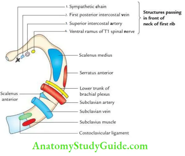
Superior view of first rib showing special feature:
- Sympathetic chain
- First posterior intercostal vein
- Superior intercostal artery
- First thoracic nerve (ventral ramus)
Note: The structures pass in front of the neck of the first rib.
Mnemonic: Chain pulls VAN. Here C of chain stands for the sympathetic chain.
Question 9. Write a short note on the sternal angle (angle of Louis).
Answer:
Sternal angle:
It is a horizontal bony angulation formed at the junction of the manubrium and the body of the sternum. It can be palpated in a living person as a bony transverse ridge about 5 cm below the suprasternal notch.
Anatomical events taking place at the sternal angle are:
Articulates on either side with the costal cartilage of the 2nd rib which helps in counting the ribs in clinical practice
- Ascending aorta ends at this level
- The Arch of the aorta begins and ends at this level
- Descending aorta begins at this level
- The pulmonary trunk divides into two pulmonary arteries at this level
- Azygos vein enters the superior vena cava at this level
- Trachea divides into two principal bronchi at this level
- Marks the junction between the superior and inferior mediastinum
- Marks the junction between two discontinuous dermatomes C4 (above) and T2 (below)
- The superior vena cava pierces the fibrous pericardium at this level
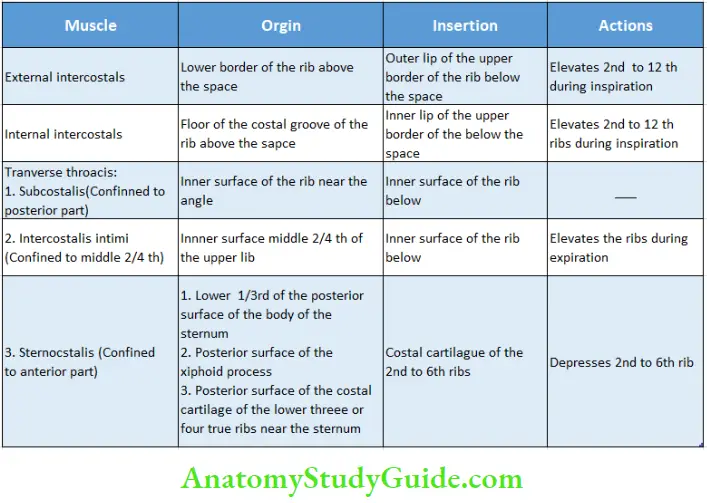
Question 10. Define intercostal space and enumerate its contents.
Answer:
Intercostal Space:
It is a space between two consecutive ribs and their costal cartilages. It extends anteriorly up to the lateral border of the sternum and posteriorly upto the body of the corresponding thoracic vertebra.
Intercostal Space Contents:
A typical intercostal space contains :
- 3 intercostal muscles.
- A neurovascular bundle.
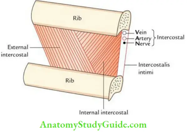
The intercostal muscles are arranged in 3 layers. From superficial to deep, these are:
- External intercostal
- Internal intercostal
- Transversus thoracic
Note:
- The transversus thoracis is divided into 3 parts: sternocostal, intercostalis intimi, and subcostalis.
- The neurovascular bundle consists of the intercostal nerve, intercostal vein, and intercostal artery. The neurovascular bundle runs between the middle and inner layers of intercostal muscles and lies in the subcostal groove on the inner surfaces of the rib near its lower border.
Question 11. Give the direction of fibers of intercostal muscles.
Answer:
These are as follows:
The direction of fibers of intercostal muscles:
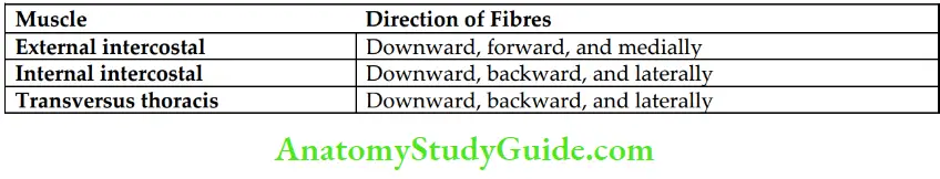
Note: The intercostal muscles are supplied by the intercostal nerves.
Question 12. Give the origin, insertion, and actions of intercostal muscles in tabular form.
Answer:
These are given in Table:
Origin, Insertion, and Actions of Intercostal Muscles:
Question 13. Write a short note on the intercostal nerves.
Answer:
Intercostal nerves:
The intercostal nerves are the anterior primary rami of thoracic spinal nerves and are located in the intercostal spaces. Thus, there are 11 intercostal nerves in the thoracic wall.
Intercostal Nerves Unique feature:
The intercostal nerves retain their segmental character, unlike the anterior primary rami of other regions where they form nerve plexuses, viz. cervical, brachial, lumbar, and sacral.
Intercostal Nerves Classification:
They are divided into two types.
- Typical intercostal nerves: They remain confined within the respective intercostal spaces of the thoracic wall, viz. 3rd, 4th, 5th, and 6th intercostal nerves.
- Atypical intercostal nerves: They extend beyond the thoracic wall, e.g., 1st, 2nd, 7th, 8th, 9th, 10th, and 11th intercostal nerves.
Note: The 7th to 11th intercostal nerves are called thoracoabdominal nerves as they leave the thoracic wall to supply the anterior abdominal wall.
Question 14. Write a short note on a typical intercostal nerve.
Answer:
The anterior primary rami of the 3rd to 6th thoracic spinal nerves are termed typical intercostal nerves and supply the muscles of the corresponding intercostal space and skin of the thoracic wall.
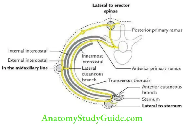
Typical Intercostal Nerve Course:
Each intercostal nerve emerges through the respective intervertebral foramen and enters into the respective intercostal space.
The courses in the intercostal space are as follows:
- In the posterior part of the intercostal space, it runs between the pleura and posterior intercostal membrane as far as the angle of the rib.
- Thereafter, it continues its course in the costal groove between the internal intercostal and intercostalis intima muscles.
- Finally, it runs between the internal intercostal and sternocostal muscles.
- At the anterior end of the intercostal space, the nerve passes in front of the internal mammary artery and runs forward, piercing the internal intercostal muscle, and anterior intercostal membrane to become the anterior cutaneous nerve.
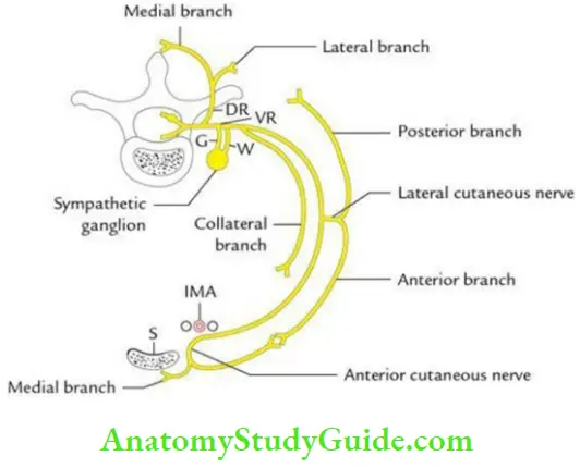
Typical Intercostal Nerve Branches:
- White ramus communicates to the sympathetic trunk.
- Collateral branch: It arises in the posterior part of the intercostal space and runs along the inferior margin of the space along the upper border of rib below.
- Lateral cutaneous branch: It appears in the midaxillary line and divides into anterior and posterior branches.
- Anterior cutaneous nerve (terminal branch) appears on the side of the sternum and divides into medial and lateral branches.
- Muscular branches.
Typical Intercostal Nerve Applied Anatomy:
- Irritation of intercostal nerves causes severe pain, which is referred to the front and side of the chest.
- Pus from tubercular abscess/cold abscess of the vertebral column tracks around the thoracic wall along the intercostal neurovascular bundle and points on the surface of the thoracic wall at 3 sites of exit of posterior, lateral, and anterior cutaneous branches of the spinal nerves.
Question 15. Describe in brief the arterial supply of the thoracic wall.
Answer:
The thoracic wall is richly supplied by blood by the intercostal arteries.
The intercostal arteries are divided into two groups:
- Anterior intercostal arteries and
- Posterior intercostal arteries.
Each intercostal space contains three intercostal arteries: one posterior and two anterior.
Anterior intercostal arteries:
- There are two anterior intercostal arteries in each intercostal space (except in the last 2 intercostal spaces).
- In the upper six intercostal spaces, they arise from the internal mammary artery.
- In the 7th, 8th, and 9th intercostal spaces, they arise from the musculophrenic artery.
- In the 11th and 12th intercostal spaces, there are no anterior intercostal arteries.
Posterior intercostal arteries:
- There is only one intercostal artery in each space, giving a collateral branch that runs parallel to it.
- The two anastomose with the corresponding anterior intercostal arteries.
- In the upper two spaces, they arise from the superior intercostal artery.
- In the 3rd to 11th intercostal spaces, they arise from the descending thoracic aorta.
Question 16. Write a short note on the internal thoracic (mammary) artery.
Answer:
Thoracic artery:
There are two internal thoracic arteries, deep to the anterior chest wall, one on either side of the sternum.
The details are as follows :
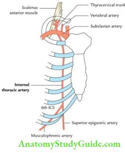
Thoracic Artery Origin:
From the first part of the subclavian artery, opposite to the thyrocervical trunk, about 2.5 cm above the medial end of the clavicle.
Thoracic Artery Course:
- First, it runs downward, forward, and medially behind the medial end of the clavicle and first costal cartilage, in front of the pleura.
- Subsequently, it runs vertically downward behind the upper six costal cartilages, 1.25 cm away from the margin of the sternum.’
Thoracic artery Termination:
It terminates in the 6th intercostal space, by dividing into superior epigastric and musculophrenic arteries.
Thoracic artery Branches:
These are:
- Pericardiophrenic artery: A long slender branch that runs along the phrenic nerve to supply the pleura and pericardium.
- Anterior intercostal arteries: The two anterior intercostal arteries are given to each of the upper six spaces.
- Perforating arteries: These accompany the anterior cutaneous nerves. The perforating branches of the 2nd, 3rd, and 4th spaces are quite large and supply the mammary gland in females.
- Superior epigastric artery: It runs downward to enter the rectus sheath behind the rectus abdominis muscle.
- Musculophrenic artery: It runs downward and laterally behind the 7th, 8th, and 9th costal cartilages. It gives rise to anterior intercostal arteries to the 7th, 8th, and 9th spaces.
- Mediastinal arteries: They are small inconstant branches that supply the thymus, front of the pericardium, and anterior mediastinal fat.
Thoracic artery Applied anatomy:
- The internal thoracic artery is sometimes ligated in the 3rd intercostal space to enhance/increase the blood supply to the heart.
- The internal mammary artery may be used for coronary artery bypass surgery also known as coronary bypass graft surgery (IMA Graft), since it is less prone to develop atherosclerosis because of its histological peculiarity.
Question 17. Give a brief account of the intercostal veins.
Answer:
Each intercostal space contains 3 intercostal veins (ICVs): two anterior and one posterior.
Anterior intercostal veins:
They are two in each upper 9 intercostal spaces. The veins of the upper 6 spaces drain into the Internal thoracic vein while those in the lower 3 spaces drain into the Musculophrenic vein.
Posterior intercostal veins:
There is only one posterior intercostal vein in each space. They drain as follows
- On the right side:
- 1st drains into the right brachiocephalic vein.
- 2nd, 3rd, and 4th unite to form the right superior intercostal vein, which drains into the arch of the azygos vein.
- 5th to 11th drain into the azygos vein.
- On the left side:
- 1st drains into the left brachiocephalic vein.
- 2nd, 3rd, and 4th unite to form the left superior intercostal vein, which drain into the left brachiocephalic vein.
- 5th to 8th drain into the accessory hemiazygos vein.
- 9th to 11th drain into the hemiazygos vein.
Question 18. Give a brief description of the mechanism of external respiration.
Answer:
Respiration consists of two components:
- Inspiration and
- Expiration.
1. Inspiration Mechanism:
- During inspiration, the volume of the thoracic cavity increases to create a negative intrathoracic pressure. As a result, the air is sucked into the lungs.
- The increase in volume of the thoracic cavity occurs due to an increase in transverse, anteroposterior, and vertical diameters of the thoracic cavity.
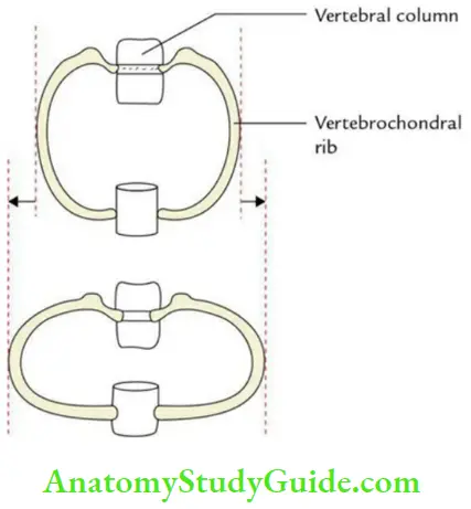
An increase in the transverse diameter of the thoracic cavity:
- Occurs due to bucket handle movements of the 7th–10th ribs (vertebrochondral ribs) mainly
- The ribs articulate in front with the costal cartilages of ribs above and behind with the vertebral column.
- Because the ribs curve downward as well as forward around the chest wall, they resemble bucket handles.
- Therefore, when the ribs are raised like bucket handles, the transverse diameter of the thoracic cavity is increased
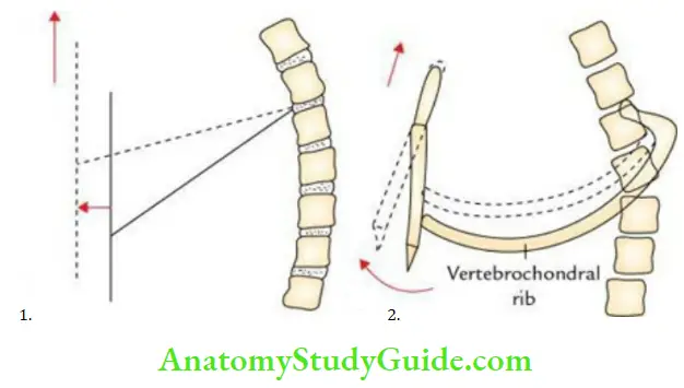
Increase in the anteroposterior diameter of the thoracic cavity:
- Occur due to pump handle movements of the vertebrosternal ribs.
- The anterior ends of the ribs lie at a lower level than the posterior ends; therefore, during the elevation of ribs, the anterior ends move upward and forward, leading to an increase in the anteroposterior diameter of the thoracic cavity.
- Since the anterior ends of vertebrosternal (the 2nd to 6th) ribs are fixed with the sternum, the sternum also moves up and down along with up and down movements of the ribs.
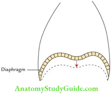
An increase in vertical diameter: Occurs due to the descent of the diaphragm as a result of its contraction.
Mechanism of expiration:
It occurs due to:
- Relaxation of the diaphragm to decrease the volume of the thoracic cavity.
- Rib muscles relax to make the rib cage smaller.
- Elastic recoil of the lung to increase the intrapulmonary pressure.
- As a result of the above mechanisms, the air is forced out.
Leave a Reply