Amalgam Restorations On Primary Teeth Introduction
Amalgam has been used as a dental restorative material for several decades. It is indicated for occlusal cavities, proximal cavities, and occlusal proximal cavities in primary molars.
Table of Contents
The cavity should be prepared in a biomechanically acceptable form to receive and retain the amalgam restoration. The outline form of the cavity preparation for amalgam restoration is based on a mandate, ‘extension for prevention’.
Read And Learn More: Paediatric Dentistry Notes
This means that an extension of the cavity into susceptible fissures is necessary for the prevention of secondary caries. The resistance form should enable the tooth and restoration to withstand masticatory forces.
A flit pulpal flor and a uniform depth of 1.5 mm are necessary for resistance even if the carious lesion is not so deep. The retention form involves mesial and distal dovetails that enable the restoration to resist displacement.
Convenience form allows adequate visibility and accessibility for restoring the cavity. The cavity preparation and restoration for amalgam class 1 and class 2 cavities in primary molars have been discussed in this chapter.
Amalgam Restorations On Primary Teeth Advantages And Disadvantages
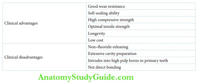
The incidence of dental caries is high in children. As amalgam restoration does not directly bond to the tooth structure, chances for microleakage or secondary caries are higher initially.
With time, the silver in the amalgam material forms an oxide layer at the margins of the restoration. The oxide layer decreases microleakage. This property of amalgam restorations is described as the self-sealing ability.
The other advantages and disadvantages of amalgam restoration are depicted in are depicted.
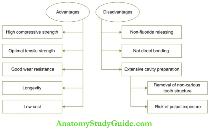
The following are the disadvantages of using amalgam, especially in restoring primary teeth:
Amalgam restorations do not contain fluoride ions, which are anti-cariogenic. There are more chances of secondary caries when non-fluoride–releasing amalgam restorations are indicated in children.
The outline form and retention form demand extensive cavity preparation. This results in the removal of non-carious tooth structures.
The resistance form of cavity preparation requires a uniform depth and a flat pulpal floor. The depth of the cavity is a concern in primary teeth as the primary pulp horns are very high.
Despite these disadvantages, amalgam is used as a restorative= material due to its superior mechanical properties, clinical longevity, and economic viability.
Class 1 Amalgam Restorations On Primary Molars
Class 1 cavity refers to the cavity in the occlusal surfaces of molars, cingulate of anterior teeth, and occlusal one-third of the buccal and lingual (palatal) surfaces of molars.
However, the cingulate of anterior teeth and buccal and lingual pits and grooves are best restored with plastic restorative materials.
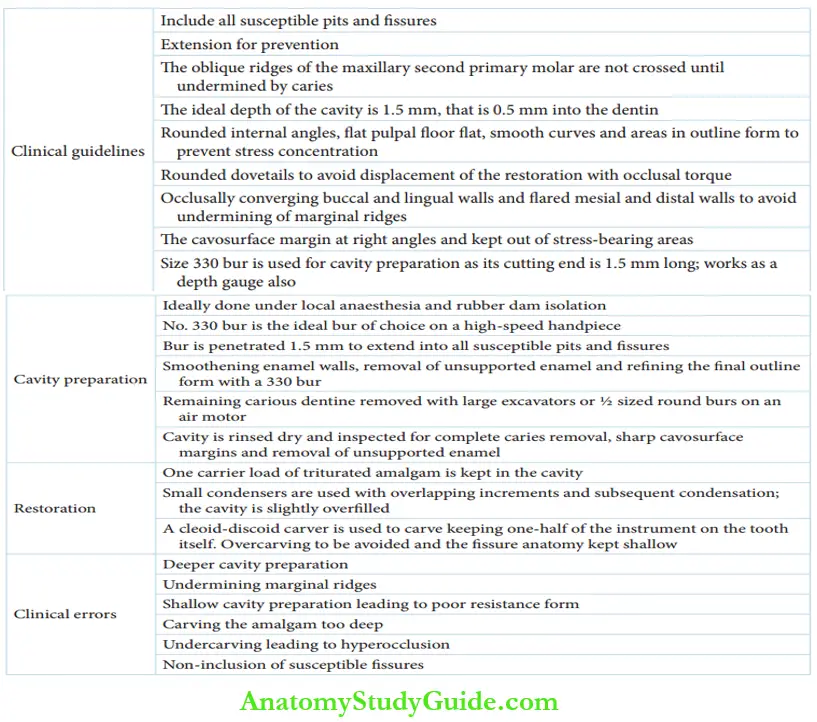
Clinical Guidelines For Class I Cavity Preparation For Amalgam Restoration
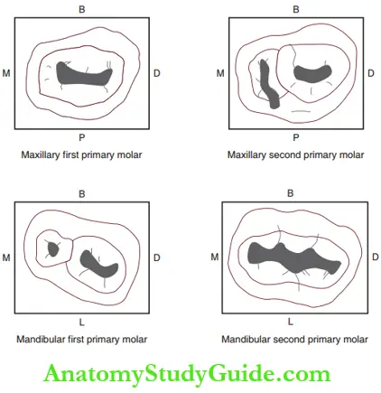
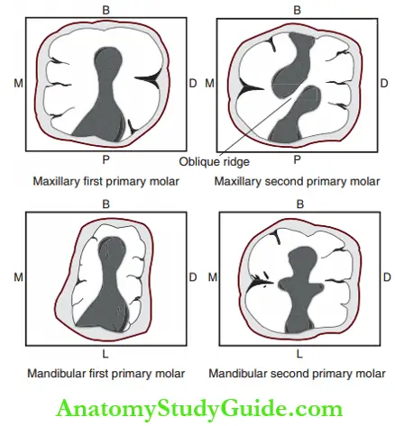
depicts the outline form of the cavity preparation for amalgam restorations. The outline form of the maxillary and mandibular primary molars has been shown.
The following are the clinical guidelines for class 1 cavity preparation for amalgam restoration in primary molars.
- The cavity outline should include all susceptible pits and fissures even if they are non-carious. This is called an extension for prevention.
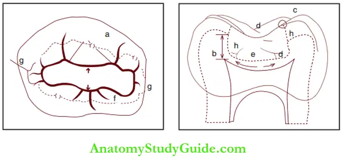
- The ideal depth of the cavity is 1.5 mm, that is 0.5 mm, into the dentin. Size 330 bur is used on a high-speed handpiece. It is the ideal choice for cavity preparation as its cutting end is 1.5 mm long. The bur works as a depth gauge also.
- The cavo surface margin should be at the right angle and kept out of stress-bearing areas.
- All internal angles have to be round, the pulpal floor flat and the outline form should have smooth curves and areas. All these features prevent stress concentration.
- Rounded dovetails avoid displacement of the restoration with occlusal torque.
- Buccal and lingual walls should converge occlusal and mesial and distal walls should fire out to avoid undermining marginal ridges.
- The oblique ridges of the maxillary second primary molar are not crossed until they are undermined by caries.
- Carious buccal developmental pits on the mandibular second primary molar have to be restored with a round teardrop-shaped restoration.
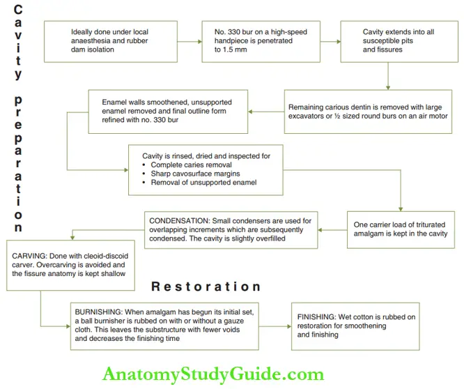
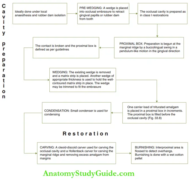
summarises the steps in class 1 cavity preparation and amalgam restoration in the form of a flowchart.
Amalgam restoration with pit and fissure sealants can be indicated as an alternative to extensive cavity preparations. The area of decay is excavated and the cavity pertains to the carious tooth structure alone.
There is no extension into the pits and fissures. The prepared cavity is restored with amalgam. Then the pit and fissure sealants are applied to the susceptible fissures.
Common Errors In Class I Amalgam Restoration On Primary Teeth
- Deeper cavity preparation may lead to pulpal exposure or involvement.
- The undermining of marginal ridges results in a poor resistance form of restoration.
- Shallow cavity preparation leads to a poor resistance form of restoration.
- Carving the amalgam too deep leaves a thin shelf of amalgam at the cavo surface margin and reduces the bulk of restoration in the stress-bearing areas.
- Undercarving leads to hyper occlusion and associated tenderness of the tooth and the temporomandibular joint.
- Non-inclusion of susceptible figures enhances susceptibility to secondary decay.
Class 2 Amalgam Restoration On Primary Molars
Class 2 cavity refers to a cavity on the proximal side of molars with or without the involvement of the marginal ridge. A class 2 cavity preparation is an extension of
the class 1 cavity preparation into the proximal surface.
The occlusal portion is called the occlusal cavity and the proximal portion is called the proximal box. depicts the outline form of the class 2 cavity preparation for amalgam restorations.
The outline form of the maxillary and mandibular primary molars has been shown.
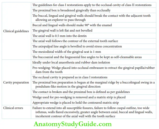
Clinical Guidelines For Class 2 Cavity Preparation For Amalgam Restoration
The clinical guidelines for class 2 restorations apply to the occlusal preparations of the class 2 restorations. The clinical guidelines and the steps of class 2 cavity preparation for amalgam restorations are illustrated respectively.
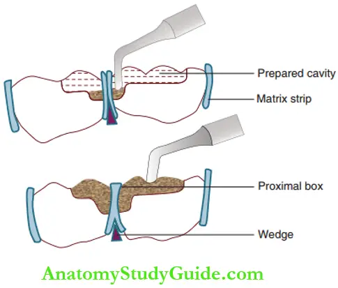
- The proximal box is broadened gingivally than occlusal.
- The buccal, lingual, and gingival walls should break the contact with the adjacent tooth allowing an explorer to pass through and through without hindrance.
- Buccal and lingual walls should be at a right angle to the enamel.
- The gingival wall is flat and not beveled.
- The axial wall should be 0.5 mm into the dentin.
- The axial wall follows the contour of the external tooth surface.
- The axiopulpal line angle is beveled to avoid stress concentration.
- The mesiodistal width of the gingival seat is 1 mm.
- The buccoaxial and the linguoaxial line angles have to be self-cleansable; hence, they appear flat out from the axial surface.
Common Errors In Class 2 Amalgam Restorations On Primary Teeth
- Failure to extend into all susceptible fissures or insufficient removal of caries enhances the possibility of secondary decay.
- Failure to follow the cuspal outline places margins of restorations under high–stress-bearing areas.
- Isthmus cut too wide can lead to a fracture of the proximal box.
- The excessive flare of the cave surface margin leads to marginal failure in the proximal box.
- Increased firing of walls can lead to a probable fracture of the proximal box.
- The decreased firing of walls keeps cavo surface line angles of the proximal box in non–self-cleansable areas (within the contact regions) increasing chances of secondary decay.
- A greater angle established between axial, buccal, and lingual walls results in a poor resistance form of the tooth.
- The contour of the axial wall that is not in line with the contour of the tooth surface also compromises the resistance form.
- The following errors cause fracture at the isthmus of the class 2 amalgam restoration:
-
- Restoration being high in occlusion
- Insufficient bulk of amalgam in the isthmus or overcarving of restoration
- Too shallow preparation at the isthmus region
Amalgam Restorations On Primary Teeth Summary
1. Amalgam as a restorative material
2. Class 1 amalgam restorations
3. Class 2 amalgam restorations
Leave a Reply