Anatomy of the Bone and Bone Composition
What are Howship’s lacunae?
Table of Contents
Howship’s Lacunae:-
:-1. There are shallow pits present on the resorbed surfaces of bone. These pits are called Howship’s lacunae. They are occupied by osteoclasts.
2. Lysosomal enzymes released by osteoclasts erode these depressions.
3. The part of the cell facing the lacunae is thrown into finger-like processes which interdigitate with similar reciprocal processes in the bone. This end of the cell is called a ruffled membrane. They are filled with vacuoles. The area surrounding the membrane, close to the bone is called the colour zone. The cells are rich in lysosomes and contain bone debris. They arise from the mononuclear cells of the bone marrow.
Read And Learn More: Anatomy Notes And Important Question And Answers
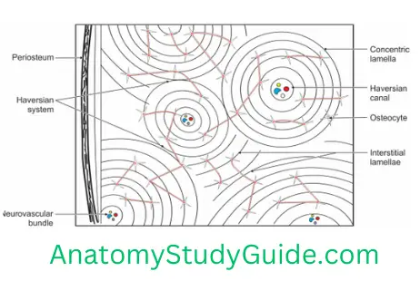
What is osteon? Or the Haversian system?
Osteon:-
- It consists of the central Haversian canal which conducts vessels, lymphatics and nerves.
- It is present only in compact bone.
- There are 18–50 concentric lamellae.
- The spaces between lamellae are called lacunae. They contain osteocytes.
Canaliculi: These are fine channels containing cytoplasmic processes of osteocytes.
State the different types of lamellae in the bone
Different Types Of Lamellae In The Bone:-
There are three types of lamellae.
1. Concentric lamellae are present around the Haversian canal.
2. Interstitial lamellae are present between two Haversian systems. They do not belong to any Haversian system.
3. Circumferential lamellae: These are of two types.
- Outer circumferential around the bone.
- Inner circumferential.
What is Volkmann’s Canal?
Volkmann’s Canal:-
It connects the Haversian system to the periosteum. It pierces lamellae and runs transversely.
State the cells of the bone. Describe each briefly.
Cells Of The Bone:-
1. Structure
The following cells are present.
1. Osteogenic cells
- They are precursors of osteoblasts.
- They are present in the cellular layer of the periosteum.
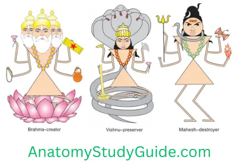
Note: According to Hindu mythology, there are three Gods. They are Brahma, Vishnu and Mahesh. The God Brahma is creator of the world, Vishnu is preservor and Mahesh is the destroyer. The simile can be used to understand the functions of bone cells. Osteoblast is creator of bone, the osteocyte is the preserver and the osteoclast is the destroyer of bone.
2. Osteoblasts
- They are active, large and basophilic cells. They are present during bone formation.
- The nucleus is round and eccentric.
3. Osteocytes
- Present in lacuna of fully formed bone.
- They have cytoplasmic processes.
4. Osteoclasts: They are
1. Bone modelling cells.
3. Formed by giant cells.
4. Multinucleated and eosinophilic.
What is osteoclast?
Osteoclasts are:-
1. Bone modelling cells.
2. Formed by giant cells.
3. Multinucleated and eosinophilic.
Compact Bone
Compact Bone Introduction: It is a specialized connective tissue with mineralized matrix.
1. Compact Bone Structure
The following cells are present.
1. Osteogenic cells are
- Precursors of osteoblasts.
- Present in cellular layer of periosteum.
2. Osteoblasts
- They are active, large and basophilic cells.
- They are present during bone formation.
- The nucleus is round and eccentric.
3. Osteocytes
- Present in fully formed bone in lacunae.
- They have cytoplasmic processes.
d. Osteoclasts are
- Bone modelling cells.
- Formed by giant cells.
- Multinucleated and eosinophilic.
2. Compact Bone Matrix: Following elements are present in matrix.
1. Organic elements
- Dense bundle of collagen fibres,
- Ground substance,
- Hyaluronic acid, and
- Protein polysaccharides.
2. Inorganic elements
- Calcium phosphate (Ca3(PO4)2)
- Calcium carbonate (CaCO3)
- Calcium chloride (CaCl2) and
- Magnesium chloride (MgCl2)
3. Osteon (Haversian system)
1. Osteon Types
- Primary osteon represents the initial structural unit of compact bone. It is formed by deposition of successive lamellae of bone. They are deposited on the walls of vascular channels.
- Secondary osteon: As the diaphysis increases in girth, all the original woven bone and primary osteon bone are removed by endosteal erosion. It undergoes a change in character. It results in development of new tracts of bone. This is called secondary osteon.
2. Osteon Features
1. It consists of the central Haversian canal which conduct vessels, lymphatics and nerves.
2. Nerve is present only in compact bone.
3. There are 18–50 concentric lamellae.
4. The spaces between lamellae are called lacunae. They contain osteocytes.
5. Canaliculi: These are fine channels containing cytoplasmic processes of osteocytes.
3. Osteon Lamellae: There are three types of lamellae.
- Concentric lamellae are present around the Haversian canal.
- Interstitial lamellae are present between two Haversian systems. They do not belong to any Haversian system.
- Circumferential lamellae: These are of two types. 1. Outer circumferential lamellae are present around bone and 2. Inner circumferential.
4. Osteon Periosteum: It has two layers
- Outer: Fibrous which contains collagen fibres.
- Inner: Cellular layer contains osteocytes.
5. Osteon Sharpe’s fibres enter from periosteum to outer circumferential lamellae.
6. Osteon Volkmann’s canal: It pierces lamellae and connects Haversian system to periosteum.It runs transversely.
7. Osteon Site
- Shaft of long bone.
- Outer layer of all compact bones.
8.Osteon Blood supply of compact bone is by nutrient artery. Osteocytes get nutrition from nutrient artery.
Leave a Reply