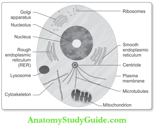
Normal Cell Structure And Functions
- A cell is a fundamental unit of life
- All cells are tridimensional, although when viewed under the light microscope on a glass slide, they appear to be flt
- Classification of cells: Except for some cells (for example, RBCs or squamous cells may lose their nucleus in the fial stages of their life cycle) all human cells have a distinct nucleus.
All nucleated cells are classified as:
Table of Contents
- Eukaryotic cells (from Greek, Karion = kernel, nucleus): DNA is enclosed by a membrane forming a distinct nuclear structure.
- Prokaryotic cells: These are primitive cells (for example, Bacteria) in which the DNA is present in the cytoplasm but is not enclosed by a membrane as a distinct nuclear structure.
Read And Learn More: Pathology for Dental Students Notes
Components of the Cell
All cells share fundamental structural components. Each cell has three essential
components:
- Cell membrane
- Cytoplasm and
- Nucleus.
1. Cell Membrane/Unit Membrane:
- It is the outer boundary of the cell
- It facilitates and limits the exchange of substances between the cell and its environment
- Light microscopy: Th membrane appears at the periphery as a thin condensation
- Electron microscopy: It is composed of three layers, each about 25 nm thick. The inner and the outer dense (electron opaque) layers are separated by a wider lucent central layer
- Biochemical components: The cell membrane is composed of a mixture of phospholipids, glycolipids, cholesterol (adds structural rigidity to the cell membrane), proteins of various sizes, and carbohydrates
- Function of cell membrane: The cell membrane plays a critical role in virtually every aspect of cell function.

2. Cytoplasm:
- Cytoplasm or cytosol: Cytoplasm or cytosol is the component of the cell, located between the nucleus and the cell membrane
- Light microscopy: Various products of cell metabolism may be seen in the cytoplasm, and appear as granules or vacuoles.
Cytoplasmic Organelles: Ultrastructurally, the cytoplasm is composed of:
- Organized cell components, or organelles,
- The cytoskeleton, and
- a cytoplasmic matrix.
These are:
- Organized components of the cytoplasm: They mainly consists of:
- Membranous systems
- Ribosomes
- Mitochondria,
- Lysosomes, and
- Centrioles.
1. Membranous System: It is composed of: The endoplasmic reticulum, and b) the Golgi complex.
Endoplasmic reticulum:
- Endoplasmic Appearance: It is a closed system of unit membranes forming tubular canals and flattened sacs or cisternae. It subdivides the cytoplasm into a series of compartments
- Endoplasmic Types: Th membranes of the endoplasmic reticulum may be “rough,” i.e. rough endoplasmic reticulum (RER) [i.e. covered with numerous attached granules composed of ribonucleic acid (RNA) and proteins], or “smooth” i.e. smooth endoplasmic reticulum (SER) (i.e. free of any particles)
- Endoplasmic Function: Rough endoplasmic reticulum is abundant in metabolically active cells.
Golgi complex:
- Golgi complex appearance: It consists of a series of parallel, doughnut-shaped flt spaces or cisternae and spherical or egg-shaped vesicles demarcated by smooth membranes
- Golgi complex Location: In epithelial cells with secretory function, it is usually located between the nucleus and the luminal surface of the cells
- Golgi complex Function: They synthesize and package cell products for the cells’ own use and for export.
2. Ribosomes:
They are found in all cells
- Ribosomes Appearance:
- They are submicroscopic particles composed of RNA and proteins in approximately equal proportions
- Two types of ribosomes namely free and attached
- Ribosomes Functions:
- Free ribosomes produce proteins for the cell’s own use
- Attached are ribosomes that produce proteins for export.
3. Mitochondria
They are present in all eukaryotic cells
Mitochondria Appearance :
- Mitochondria are small, usually elongated structures that vary in size and configuration according to the nutritional status of an organ
- Each mitochondrion is composed of two membranes. The outer membrane is a continuous, closed-unit membrane.
- Running parallel to the outer membrane is the inner membrane that forms numerous crests or invaginations.
Mitochondria of Function:
- Most important function is the formation of energy-producing adenosine triphosphate (ATP) from phosphorus and adenosine diphosphate (ADP).
- ATP is exported into the cytoplasm and is an essential source of energy for the cell
- Mitochondria possess their own DNA that is independent of nuclear DNA.
4. Lysosomes:
Lysosomes are intracellular organelles that contain degradative enzymes
Lysosomes Appearance:
Electron microscopically, lysosomes appear as spherical or oval structures of heterogeneous density and variable diameter
Lysosomes Function:
- Lysosomal enzymes perform the digestion of a wide range of macromolecules, including proteins, polysaccharides, lipids, and nucleic acids.
- They participate in the removal of phagocytized foreign material. Play an important role in certain storage diseases (lysosomal storage disorders).
5. Centrioles:
Centrioles Appearance: They are short tubular structures, usually located in the vicinity of the concave face of the Golgi complex
Centrioles Function: Play a key role during cell division.
1. Cytoskeleton:
The skeleton of the cells, and hence, the ability of cells to maintain a particular physical shape, maintain polarity, movement of cells, and organize the relationship of intracellular organelles is provided by a family of fibrillar proteins This intracellular scaffolding of fibrillar proteins is called the cytoskeleton
Types of Cytoskeleton: In eukaryotic cells, there are three major types of cytoskeletal proteins: Actin filaments (microfilaments, tonofiaments), intermediate filaments, and microtubules.
- Actin microfilaments: They interact with many other proteins that regulate their length
- Electron microscopy: They appear as bundles of longitudinal cytoplasmic figments crisscrossing the cytoplasm and often converging on specific targets such as desmosomes
- Functions: Actin microfilaments perform several essential functions within cells as linkage filaments coordinating the activity of divergent cell components.
- Intermediate filaments:
- Appearance: They were initially identified in electron microscopy because of their diameter (7 to 11 nm); hence, intermediate filaments (IFs). They are larger than actin microfilaments and smaller than microtubules
- Functions: Individual types of IFs have characteristic tissue-specific patterns of expression. This can be used for identifying cells of origin of poorly differentiated tumors.
- Microtubules:
- They are 25-nm-thick fails and appear as hollow, tube-like structures
- Functions: Microtubules are an integral component of cilia, flagella, and centrioles. This, they participate in cellular events requiring motion.
2. Cytoplasmic matrix of Centrioles: The space within the cytoplasm, not occupied by the membranous system, the cell skeleton, or by the organelles, is called the cytoplasmic matrix. It is composed of proteins and free ribosomes.
3. Nucleus of Centrioles:
- It is present within the cytoplasm
- Nuclear membrane: It is a smaller, approximately spherical dense structure enclosed within the nuclear membrane, or nuclear envelope
- Content: It contains mainly deoxyribonucleic acid (DNA) that governs the genetic and functional aspects of cell activity
- Nucleolus: In normal resting nuclei, the nucleoli are seen as round or oval structures occupying a small area within the nucleus.
Introduction To Pathology
Definition: Pathology is the scientific study (logos) of disease (pathos). It mainly focuses on the study of the structural and functional changes in cells, tissues, and organs in disease.
Learning Pathology:
The study of pathology can be divided into general pathology and systemic pathology.
- General pathology: It deals with the study of mechanisms, and basic reactions of cells and tissues to abnormal stimuli and to inherited defects.
- Systemic pathology: This deals with the changes in specific diseases/responses of specialized organs and tissues.
Scientific Study of Disease:
- The disease process is studied under the following aspects:
Etiology of Pathology:
- The etiology of a disease is its cause.
- The causative factors of a disease can be divided into two major categories:
- Genetic and acquired (for example Infectious, Chemical, Hypoxia, Nutritional, Physical).
- Most common diseases are multifactorial due to a combination of causes.
Pathogenesis:
It refers to the mechanism by which the causative factor/s produces structural and functional abnormalities. Pathogenesis deals with a sequence of events that occur in the cells or tissues
from the beginning of any disease process.
With the present advances in technology, it is possible to identify the changes occurring at the molecular level and this knowledge is helpful for designing new therapeutic approaches.
- Latent period: Few causative agents produce signs and symptoms of the disease immediately after exposure. Usually, etiological agents take some time to manifest the disease (for example, Carcinogenesis) and this time period is called the latent period. It varies depending on the disease.
- Incubation period: In disorders caused by infectious (due to bacteria, viruses, etc.) agents, the period between exposure and the development of disease is called the incubation period. It usually ranges from days to weeks. Most infectious agents have a characteristic incubation period.
Morphologic Changes:
All diseases start with structural changes in cells. Rudolf Virchow (known as the father of modern pathology) proposed that injury to the cell is the basis of all diseases. Morphologic changes refer to the gross and microscopic structural changes in cells or tissues affected by disease.
Gross of Morphologic :
- Many diseases have characteristic gross pathology and a fairly confident diagnosis can be given before light microscopy.
- For example, serous cystadenoma of the ovary usually consists of one cystic cavity containing a serous fluid; cirrhosis of the liver is characterized by total replacement of the liver by regenerating nodules.
Microscopy of Morphologic :
1. Light microscopy: Abnormalities in tissue architecture and morphological changes in cells can be studied by light microscopy.
- Histopathology: Sections are routinely cut from tissues and processed by pararffiembedding. The sections are cut from the tissue by a special instrument called a microtome and examined under a light microscope.
- In certain situations (for example, Histochemistry, rapid diagnosis), sections are cut from tissue that has been hardened rapidly by freezing (frozen section). The sections are stained routinely by hematoxylin and eosin.
- Pathognomonic abnormalities: If the structural changes are characteristic of a single disease or diagnostic of an etiologic process it is called as pathognomonic.
- Pathognomonic features are those features that are restricted to a single disease, or disease category. The diagnosis should not be made without them.
- For example, Aschof’s bodies are pathognomonic of rheumatic heart disease and Reed Sternberg’s cells are pathognomonic of Hodgkin’s lymphoma.
- Cytology: The cells from cysts, body cavities, scraped from body surfaces or aspirated by fine needles from solid lesions can also be studied under a light microscope. This study of cells is known as cytology and is used widely, especially in the diagnosis and screening of cancer.
- Histochemistry (special stains): Histochemistry is the study of the chemistry of tissues, where tissue/cells are treated with specific reagents so that the features of individual cells/ the structure can be visualized, Prussian blue reaction for hemosiderin.
- Immunohistochemistry and immunofluorescence: Immunohistochemistry and immunofluorescence utilize antibodies (immunoglobulins with antigen specifiity) to visualize substances in tissue sections or cell preparations. The former uses monoclonal antibodies linked chemically to enzymes and later fluorescent dyes.
2. Electron microscopy: Electron microscopy (EM) is useful to study changes at the ultrastructural level, and to the demonstration of viruses in tissue samples in certain diseases. The most common diagnostic use of EM is for the interpretation of biopsy specimen from the kidney.
Molecular Pathology:
- Most diseases can be diagnosed by the morphological changes in tissues. But, with the present advances in diagnostic pathology, diseases can be analyzed by the molecular and immunological approaches.
- Molecular pathology has revealed the biochemical basis of many diseases, mainly congenital disorders, and cancer.
- These techniques can detect changes in a single nucleotide of DNA. In situ hybridization can detect the presence of specific genes or their messenger RNA in tissue sections or cell preparations.
- Minute quantities of nucleic acids can be amplified by the use of the polymerase chain reaction. DNA microarrays can be used to determine patterns of gene expression (mRNA).
Functional Derangements and Clinical Manifestations:
- Functional derangements: The effects of genetic, biochemical, and structural changes in cells and tissues are functional abnormalities.
- For example, excessive secretion of a cell product (for example, Nasal mucus in the common cold); insufficient secretion of a cell product (for example Insulin lack in diabetes mellitus).
- Clinical manifestations: Th functional derangements produce two clinical manifestations of disease, namely symptoms and signs. Diseases characterized by multiple abnormalities (symptom complex) are called syndromes.
- Prognosis: The prognosis forecasts (predicts) the known or likely course (outcome) of the disease, and therefore, the fate of the patient.
- Complications: It is a negative pathologic process or event occurring during the disease which is not an essential part of the disease. It usually aggravates the illness.
- For example, perforation and hemorrhage are complications that may develop in typhoid ulcers of the intestine.
Leave a Reply