Chromosomes and their Aberrations
Gene
Introduction: It is the hereditary unit formed by segments of DNA (deoxyribonucleic acid).
Table of Contents
1. Function: It synthesizes the polypeptide.
2. Number: There are about 80,000 genes in a human cell.
3. Composition:
- Deoxyribose sugar
- Nitrogen bases, and
- Phosphates.
Read And Learn More: General Histology Question And Answers
4 Parts:
- The functional part is called exon.
- The silent part is called intron.
5. Position:
- Locus: It is the position of the genes on chromosomes.
- It is described in relation with the centromere.
- Alleles (allos—another): Any alternative form of a gene that can occupy a particular chromosomal locus.
6. Types:
1. Regulator gene:
- It represses (prevents) the activities of genes.
- It inhibits protein synthesis.
2. Operative gene:
1. Site: It is present at one end of particular gene.
2. Function: A gene that serves as a starting point for reading the genetic code and controls the activity of the structured genes by interacting with repressor.
3. Dominant gene: It expresses its physical or biochemical trait, when allelic genes are either homozygous or heterozygous,
Example :
- For the person showing brachydactyly, the prominent genes are NB, and BB.
- The tallness is caused by the dominant gene, the genotype of the tall individual is T: T, T: t.
4. Co-dominant gene: When both allelic genes are dominant but of two different types, both traits may have concurrent expression.
5. Recessive gene: It expresses its biochemical and physical traits only in the homozygous state, example albinism which is recessive
6. Sex-linked gene:
- Abnormal gene located on X or Y chromosome.
- ‘X’ linked inheritance is more common and is mostly expressed by recessive gene.
7. Sex limited gene: Born by autosomes but trait is expressed in one of the sex, e.g. gout, baldness.
8. Carrier gene: Heterozygous recessive gene acts as carrier gene. It may be expressed in subsequent generations.
Barr body (sex chromatin)
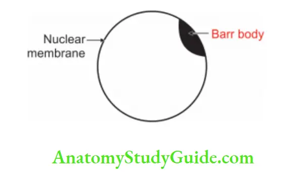
1. Introduction: It is an inactivated X chromosome attached to a nuclear membrane. It is found by Barr and Bertram in 1949.
1. Morphology:
- Site: It is attached to the nuclear membrane.
- Nature: Heterochromatin (State of chromatin in which it is dark staining and tightly coiled, forming an irregular clump or Barr bodies in the nuclei of cells in interphase.)
- Shape: Planoconvex
- Dimension: 1 μ
- Staining: Darkly stained
- In female ♀ ____________ XX
- In male ♂ ______________ (XY). There is only one X chromosome which is for cellular function.
- Y chromosome is for the determination of sex. Hence, there is no Barr body.
2. Person with XO: Will be female ♀ as the Y chromosome is absent. The only X chromosome that is present will be in an extended stage and no Barr body is seen.
The inert X chromosome (Barr body) also divides during cell division. This Barr body is also known as sex chromatin.
- Cell + Barr body = Chromatin positive
- Cell – Barr body = Chromatin negative
3. Appearance: It appears by 2nd week of gestation.
4. Lyon’s hypothesis: The number of Barr bodies is less than the total number of X chromosomes.
Number of X chromosome – 1 = Number of Barr bodies, Example.
In females ♀ with XX chromosome, there is a Barr body.
In males ♂ with XY chromosomes, there is no Barr body.
The absence of Barr body is not enough to prove that cell is from a male ♂.
Number of Barr bodies in different syndromes:

5. Applied anatomy:
- It is helpful for the determination of sex in case of genital ambiguity.
- It is a supplementary test for chromosomal aberrations.
- It is helpful for the diagnosis of various syndromes.
Structure of chromosome
1. All chromosomes consist of two parallel identical filaments called chromatids.
2. They are joined together at a narrowed constriction called primary constriction inside the primary constriction, there is a pale staining region called centromere.
3. Free ends of chromatids are called telomeres. Each chromatid is divided by a centromere in two arms.
4. In certain chromosomes, there is another narrowing (constriction) near 1 end of each chromatid called secondary constriction.
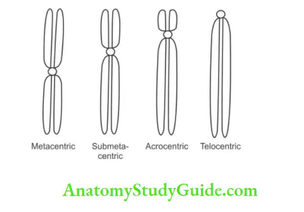
It is stained faintly. If the secondary constriction is close to the telomere, then the terminal knob of the chromosome is termed as satellite (companion). As per the position of the centromere, chromosomes are grouped in 4 types
Types and position of chromosomes:

5. The chromosome is arranged in descending order of length. The pair No. 1 is the longest and the pair No. 22 is the shortest. They are grouped into 7 groups. They are denoted as A to G.
Classes of the chromosome:

6. Sex chromosomes are X and Y, X belongs to the C group and Y belongs to the G group.
7. Each cell contains a fixed number of chromosomes, which is characteristic of that species or organism.
8. In the somatic cell (body cell) of man, the number is 46, which is a diploid number.
9. In germ cells, i.e. ova and sperms, the number is 23, called haploid number. When fertilization takes place, the union of two haploid cells restores a diploid number of the fertilized ovum. The number of sets is termed as ploidy.
10. If more than two sets are present, the cell is said to be polyploid. Chromosomes are in multiples of haploid number that is tetraploid (tetra—4) it has 4 times haploid number of chromosomes. The Triploid (tri—3) number of chromosomes will be 69, i.e. 3 times the haploid number.
Classification of chromosomes (Chrom—color, soma—body)
1. Structure: Each chromosome is made up of two identical parallel filaments called chromatids, which are held together at a narrow-constricted region, usually pale staining, known as primary constriction, or centromere or kinetochore. This structure is visible only during the metaphase stage of cell division.
1. Chromosome consists of:
- Centromere: The constricted part of a chromosome is called the centromere.
- Telomere: Free ends (arms) of the chromosome.
- Satellite body: Part distal to secondary construction.
2. Function of chromosome: The chromosome is for the perpetuation of species.
3. Classification:
According to functions:
1. Autosomes: 22 pairs in humans.
2. Sex chromosomes decide the sex of a person.
- Male ♂ _________ XY
- Female ♀ _________ XX
According to the positions of the centromere (Denver’s classification).
Positions of the Centromere:

4. Applied anatomy: Chromosomes are mapped according to the length of the arm and position of the centromere and it is called karyotyping.
Chromosomal aberrations
Introduction: It is the change in the structural component of the chromosome or the number of chromosomes. The deletion of a segment or addition of a segment from other chromosomes results in structural aberration. The change in number leads to numerical aberration.
1. Factors: The following are the factors for the chromosomal variations.
- Late age of parents for conception.
- Genes predisposing to non-disjunction.
- Viral infection during pregnancy.
- Exposure to radiation.
- Autoimmune disease of parents.
2. Types:
1. Numerical type:
- Aneuploidy: 2n + 1, 2n – 1
- Polyploidy: Multiple of n except 2n, for example, triploidy.
2. Structural type: It causes changes in the number or sequence of genes.
1. Inversion: It is the chromosomal aberration. It is caused by the inverted reunion of a chromosome segment after the breakage of a chromosome at 2 points. It results in a change in the sequence of genes or nucleotides, e.g. the sequence mnopq may be inverted to mnqop. It may be
- Paracentric: It is on one side of the centromere.
- Pericentric: It is surrounding the centromere.
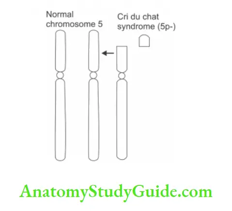
2. Deletion: A portion of the chromosome is lost, For Example. cri du chat syndrome or Philadelphia chromosome. It may be
Termination-Interstitial:
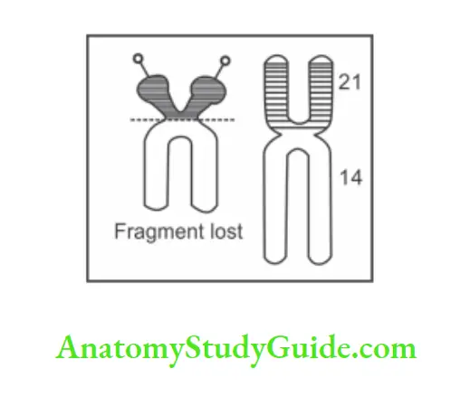
3. Translocation: It is the displacement of a portion of one chromosome to another chromosome. It is of two types.
- Robertsonian translocation, and
- Reciprocal translocation.
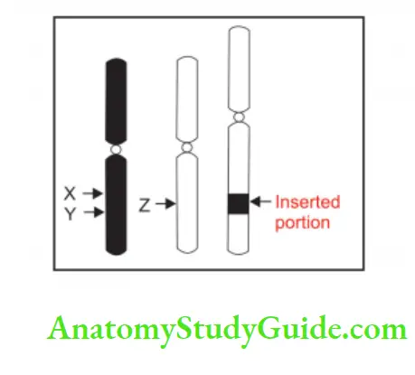
4. Insertion:
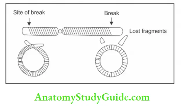
5. Ring chromosome:
6. Isochromosome:
7. Duplication:
Chromosome banding
Introduction: The chromosomes are identified by the banding technique.
1. Procedure: The chromosomes are treated 1st with trypsin and then stained. All the chromosomes are stained with dark and light regions. The dark regions are known as bands. The position of bands in normal chromosomes remains fixed and is different for different chromosomes.
2. Types of banding: Banding can be of different types.
- GTG = Giemsa Trypsin Giemsa banding.
- ASG = Acetic saline Giemsa banding.
- Q = Quinacrine mustard banding.
3. Importance of banding: It helps in
- Identification of individual chromosomes.
- Confirmation of deletion and inversion
Trisomy 21
Introduction: It is the most common autosomal abnormality syndrome described by John Langdon Haydon Down in 1866.
1. Genotype: Trisomy 21 (Down’s syndrome, Mongolism) 47 XX ( + 21) or 47 XY (+ 21).
2. Phenotype: It is the most common autosomal abnormality.
3. Incidence: 1:650 to 700 newborns.
4. Symptoms and pathophysiology: There are three chromosomes in chromosome no. 21.
1. Forms of Down syndrome: There are 3 forms
Trisomy 21 is the most common chromosomal abnormality: An extra copy of a chromosome in 21 no. of the chromosome. It may be male ♂ or female ♀
- If there is XY in 23 no. of chromosomes, it is male ♂.
- If there is XX in 23 no. of chromosomes, it is female♀.
Translocation Down syndrome: It is less common. It affects 3% of people.
Mosaic Down syndrome: It affects 2% of people.
2. Cause: Is not known.
3. It is the result:
- Non-disjunction: In 95% of cases, Down syndrome is the result of non-disjunction. Here chromosomes do not split apart. This can occur in 1st or 2nd stage.
- Robertson’s translocation: In 4% of cases, Down syndrome is the result of Robertson translocation. A chromosome number 21 gets attached to chromosome number 14.
- An abnormal chromosome is called 14, 21. Here number of chromosomes is 46.
- Mosaic: 1%—cells mixed.
5. Risk factors:
1.Major risk factors: Maternal age
- Tire incidence of Down syndrome is 1:1500 in females ♀ who are less than 20 years old.
- The incidence of Down syndrome is 1:25 in females ♀ who are more than 45 years old.
- How the maternal age affects the disjunction: In females ♀ all the X chromosomes are formed before birth.
- As the maternal age advances, the X chromosome also ages. The frequency of non-disjunction in meiosis increases with age.
- The number of aneuploidy cells increases with maternal age.
2. Minor risk factors: Chromosomal variations are
- Genes predisposing to non-disjunction.
- Viral infection during pregnancy.♂ and female
- Exposure to radiation
- Autoimmune disease of parents.
6. Clinical features:
- Effect on brain
- The affected individual is mentally retarded.
- He may suffer from Alzheimer’s disease.
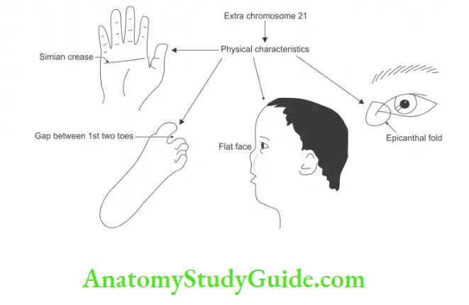
7. Effect on face:
- Flat facial profile
- The nasal bridge is flat.
- The palpebral fissure is slanting upwards at the lateral end.
- The epicanthic fold of eyes.
- Maxilla is small.
- The palate is narrow, so the oral cavity cannot accommodate the tongue.
- The tongue protrudes out of the mouth.
8. Effect on the heart: In about 50% of the cases, there is congenital heart disease. There is an atrioseptal defect.
9. Effect on the gastrointestinal tract: Duodenal atresia
10. Effect on blood: Acute lymphatic leukemia
11. Reproductive: Sterility in males ♂
12. Physical characteristics:
- Simian crease
- The gap between the two toes
13. Investigations:
1. Blood:
There is a decrease of the:
- Alpha-foetoprotein.
- Unconjugated estriol (uE3)
There is increase of the:
- Human chorionic gonadotrophin (hCG)
- Inhibin A
2. Ultrasound: Nuchal transparency—how much light passes through neck.
Turner syndrome (45 X)
Introduction: Described by Turner in 1938.
1. Monosomy 23:
1. It is not a recessive or dominant sex-linked disorder.
2. It is loss of one X chromosome
3. Incidence – 1:2000 girls
4. Aetiology
Major risk factors: Maternal age
- How the maternal age affects the disjunction: In females ♀, all the X chromosomes are formed before birth.
- As the maternal age advances, the X chromosome also ages. The frequency of non-disjunction in meiosis increases with age.
- The number of aneuploidy cells increases with maternal age.
Minor risk factors for: Chromosomal variations are
- Genes predisposing to non-disjunction.
- Viral infection during pregnancy.
- Exposure to radiation.
- Autoimmune disease of parents
2. Incidence: 1:5000 newborns.
3. Genotype: 45 X
4. Phenotype: In females Barr’s body is absent.
5. Clinical manifestations:
- Stature: Short female ♀
- Mouth: Shark-like.
- Lip: Upper— Curved, Lower — Straight.
- Chest: Shield-like— breast underdeveloped, widely placed rudimentary nipples.
- Genitalia: Small—ovarian dysgenesis infertility Most often sterile (infertile)
- In females gonads are not developed at puberty.
- Mental retardation
- It is a genetic disorder that only affects females ♀.
- But girl with a Turner’s syndrome has only 45 chromosomes.
- Face more health problems than the average female ♀.
- Low intelligence.
- Webbing of the neck.
Klinefelter syndrome (47 XXY)
Introduction: Described by Klinefelter in 1942.
1. Interesting facts:
- George Washington has this syndrome.
- One Barr body is present.
- Commonest chromosomal disorders in male QT7.
- An extra X chromosome in male Qf7.
- Women who get pregnant after the age of 35 are slightly more likely to have a boy with the syndrome than younger women.
- It is not inherited. These chromosomal changes occur as random events.
- Nondisjunction results in reproductive cell with an abnormal number of chromosomes.
2. How does Klinefelter come about:
An egg or sperm cell may gain one or more extra copies of the X chromosome as a result of nondisjunction. If one of the atypical reproductive cells contributes to the genotype of a child, the child will receive an extra X.
It is because of nondisjunction. It is the failure of paired chromosomes to disjoin (separate) during cell division so that both chromosomes go to one daughter cell and none to the other.
3. Incidence: It occurs in about 1 in 500 to 1000 baby boys. l:1000 live male ♂ birth.
4. Phenotype: Male ♂.
5. Genotype: 47 XXY
6. Signs and tests:
1. Karyotyping: A test to examine chromosomes in a sample of cells, which can help identify genetic problems. This test can count the number of chromosomes and different structures in chromosomes.
2. Semen test: It is a test to measure the amount and quality of a man’s semen. This test would be completed because the most common symptom is infertility.
7. Clinical manifestations:
- Abnormal body proportions: Long legs, short trunk. Shoulder equal to the hip size.
- Gynecomastia: Abnormally large breast.
- Infertility
- Sexual problems
- Scanty pubic hair.
- Small, firm testicles.
- Azoospermia.
8. Diagnosis:
- Prenatal diagnosis by chorionic sampling or amniocentesis in which fetal tissue is extracted and DNA is examined.
- There is no way to detect the carrier of this disorder.
- Karyotyping
- Semen test
- Amount and quality of semen
9. Complications:
- Enlarged teeth with a thinning surface (taurodontism)
- ADHD (attention deficit hyperactivity disorder)
- Breast cancer in men
- Depression
- Learning disabilities include dyslexia (a disorder involving difficulty in learning to read or interpret words, letters and other symbols).
- Osteoporosis: Bones become fragile and more likely to fracture
- Varicose veins
- Autoimmune disorder: Lupus, Rheumatoid arthritis, Sjogren syndrome
10. The extra X: chromosome has many effects on the body particularly on the testis.
- No puberty
- No testosterone
- The size of the testis is 1/8th the size.
11. Treatment:
1. No treatment is available to change a person’s chromosomal makeup.
2. Testosterone replacement for
- Muscularization
- Positive mental attitude
- Less fat on the abdomen
- Body hair
- Infertility
Cri du chat syndrome (5p-)
Cri du chat syndrome:
Cat is crying for losing heart ❤️ shaped purse having ₹5 in it.
Illustration
1. Cat is used to denote the infant’s cry which sounds like a mewing of a cat.
2. Crying for ₹5 indicates low intellectual and mental retardation.
3. Heart ❤️ shaped purse indicates congenital heart disease (VSD—ventral septal defect).
4. Losing ₹5 indicates deletion of small arm of chromosome no. 5
5. The picture has a small head indicating microcephaly.

Introduction: Cri du chat (French for the cry of cat) syndrome. It is an example of a condition caused by structural chromosomal aberration.
1. Aetiology: Genetic disorder that is caused by a missing piece of chromosome no.5
- Autoimmune disease of parents.
- Viral infection in pregnancy.
- Conception occurred late.
- Genes predisposing to nondisjunction.
- Exposure of radiation.
2. Incidence: 1:50,000
3. Gender variation: Equally in male ♂ and female ♀ No specific race.
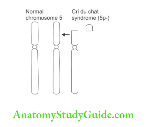
4. Genotype: It is a microscopically detectable deletion of terminal portion of short arm of p of chromosome 5 (5p-)
5. Clinical manifestations:
About brain:
- Microcephaly
- Mental retardation
- Intellectual disability
About face:
- Abnormally shaped ears
- Premature graying of hair
- Skin tags just in front of the ears and eyes
- Wide set of eyes
- Oblique palpebral fissure
- Saddle nose
- Cat-like cries during infancy.
- Excessive drooling (dribbling of saliva)
Larynx malformed: High-pitched voice
General features:
- Low birth weight and slow growth.
- Muscular hypotonia.
- Partial webbing or fusing of fingers or toes
- Single line on the palm of the hand
- Slow or incomplete development of motor skills
Behavioral problems, constipation, and Feeding problems.
6. Diagnosis: Possible to detect it with amniocentesis, chorionic villus sampling, or CVS
7 Treatment: No ways to manage the symptoms
- Speech therapy
- Physical therapy
- Special education
8. Fun facts:
- Less noticeable as the baby gets older.
- Geneticist
- The main issue located in band 5P 15.2
- Not dominant or recessive trait
- 80% of the time defective chromosomes from the father’s side.
- It happens randomly.
- It is not hereditary.
- Lifespan Normal
Nondisjunction
Introduction:
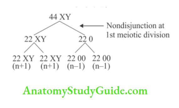
1. Failure of normal migration of chromosomes or chromosomes during anaphase of meiosis I.
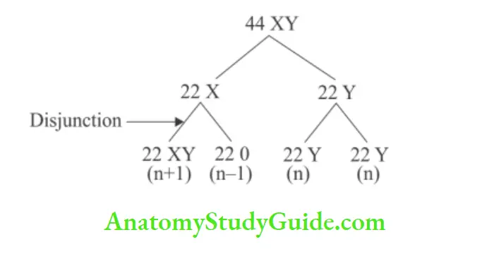
2. Failure of migration of chromatids or chromatids during meiosis II.
Reason: Factors responsible
- Faulty spindle formation.
- Slow movement of chromatid or chromosome during anaphase.
- Radiation, viruses, and autoimmune diseases, for example. myasthenia gravis, AIDS.
It results in: the formation of abnormal gametes
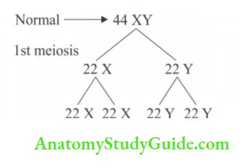
Normally each sperm has a haploid number.
1. In nondisjunction:
2. Nondisjunction: At 2nd meiotic division
3. Change in number of chromosomes: It can occur in sex chromosomes and autosomes when (n + 1) fertilized by n
→ Result 2n + 1 chromosomes (trisomy) (n-1) fertilized by n
→ (2n-1) chromosomes (monosomy)
Effect of fertilization—abnormal gamete.
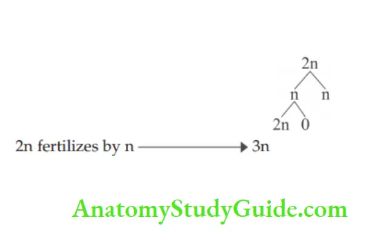
In meiosis II, if all chromatids go to one side. One daughter cell with 2n and other dies
Aneuploidy (An —not, eu—good, ploidy—multiplication)
If number of chromosomes in body cell is either
1. More than a diploid number: But not multiple of haploid number, for example. 47 chromosomes.
2. Less than diploid number: But not haploid number,
For example:
1. An abnormal X chromosome
2. One more autosome goes in other cell during division
- Each of the 46 chromosomes is a member of a homologous pair.
- One member of each pair is received from the mother and one from the father.
- The members of pairs are called homologs.
- 22 pairs are similar in males ♂ and females ♀ and are called autosomes.
- Chromosomes in the remaining pairs are sex chromosomes. In female ♀ sex chromosomes (X and X) are identical so females ♀ are homogametes.
- In males ♂, one is X and the other is Y which are unequal, so males ♂ are heterogametic.
- Homologous chromosomes in each pair of autosomes are indistinguishable.
- i. e. two chromosomes forming pair number 5 are not identified separately as they appear the same.
Spectral karyotyping (Sky)
Karyotyping: Analysis of chromosomes.
1. Definition: A picture of all the chromosomes in an individual cell arranged in homologous pairs. They are sorted according to size. Autosomal chromosomes are arranged first. Sex chromosomes are arranged last. It is also called the painting of chromosomes.
2. Spectral karyotyping: A fluorescent tag (dye) is given. They are given by different colors.
Prerequisite for spectral karyotyping:
- The sequence of the genome must be known.
- DNA must be in single strand.
- Segment-wise probe is made.
- Different probes will bind different regions.
- The designing of the probe is equally important.
- Complimentary probes are designed.
- Depending upon various colors, we can identify the various chromosomes.
- Hybridization of colors is possible in swapping events. It is good for
- Translocation and
- Substitution of chromosomes
3. Disadvantage of spectral karyotyping: It does not help to find other type of mutation
Example:
- Inversion of chromosome – The changes are in the same chromosome.
- Duplication of chromosome
4. Prerequisite: The cells must be in metaphase. They are black-and-white chromosomal karyotyping.
5. There are two types of chromosomes:
1. Autosomal chromosomes: The word “soma” means body. They have influence on body characters. They are associated with the structure and function of all cells not associated with sex. There are 44 autosomal chromosomes.
2. Sex chromosomes: They determine the sex. There are two sex chromosomes.
6. Importance of karyotyping:
1. To study differences in chromosome size, shape, and structure.
2. Determination of sex
3. Diagnose chromosomal disorder.
- Mutations
- Trisomy 21—Down’s syndrome
- Sex chromosome
4. Chromosomal problems
- Trisomy: Extra copy of chromosome
- Monosomy: Missing copy of a chromosome (only 1 copy in zygote)
- Partial addition: Extra part of a chromosome
- Partial deletion: Missing part of a chromosome.
- Inversion: A piece of chromosome breaks off, flips around, and reattaches (changes the order of gene)
- Translocation: A piece of chromosome breaks off, attaches to a different chromosome
What are genetic disorders?
Introduction: The mapping of chromosomes depending on the length and position of the centromere is called karyotyping.
1. Procedure: It is done by the microculture of lymphocytes. The cells are grown in culture media phytohaemagglutinin (PHA). The cell division is arrested in metaphase by adding colchicine.
The spreads of the chromosome are counted and photographed. The images of each chromosomes are cut out and arranged as per classification.
Karyotyping is done on the basis of:
- The total length of chromosomes.
- Position of centromere.
- The relative length of two arms.
- Banding pattern.
- The chromosomes are arranged according to their length in descending order.
- Identical chromosomes are paired in karyotyping.
- Then chromosomes paired are numbered 1 to 22 in descending order of length. i.e. pair No.1 is long, pair No. 22 is short. They are grouped into 7 groups.
- They are noted as A to G. The chromosomes are placed separately.
Classes of chromosomes:

2. Karyotyping helps to:
Identify patterns of the abnormal chromosomes.
Determination of the sex.
Leave a Reply