Development Of Occlusion Introduction
Occlusion can be described as the maximum interdigitation of the opposing full complement of teeth.
Table of Contents
A proper interdigitation of teeth is essential for efficient mastication. Teeth erupt in the alveolus and move Inman in the occlusal direction until contact is established with the opposing teeth. When a tooth erupts and reaches the occlusal plane, the point of initial contact with the opposing tooth plays a role in deciding its final position in occlusion.
The initial contact of the erupting tooth on the slope of the cusp of the opposing tooth slides the tooth down the inclined cuspal slope. This cuspal inclination guides a proper settling of occlusion into a cusp-to-occlusal fossa relationship, enabling maximum contact of the surface area. The development of such occlusion occurs in a staged manner as described below.
Read And Learn More: Paediatric Dentistry Notes
Stages Of Dental Development
With postnatal growth and development of the craniofacial skeleton, the teeth and alveolar process undergo strategic, timed maturation. This can be distinctly identified in four stages:
- Predentate stage: Before the eruption of the primary teeth, the gum pads of the maxillary and mandibular arches exist in a particular relationship with each other, enabling suckling action. This stage extends from birth to 6–7 months of age.
- Deciduous dentition stage: This stage, extending from 6–7 months to 6–7 years of age, is when only the deciduous teeth are present in the oral cavity. The deciduous teeth provide an adequate occlusion required for this age.
- Mixed dentition stage: Th increase in masticatory demand of growing age is met by the gradual replacement of deciduous dentition. A smooth transition occurs from 6–7 to 12–13 years of age. During this period, the larger permanent teeth erupt one by one to replace the exfoliated predecessor in a sequential manner.
- Permanent dentition stage: With all primary teeth replaced with permanent teeth, the dentition matures from being a mixed dentition to a permanent dentition. This stage extends from around 13 years of age and the permanent teeth are expected to last for a lifetime.
Predentate/Neonatal Stage
The dentate stage extends from birth till 6–7 months of age when the first primary tooth erupts. The dentate maxillary and mandibular jaws that represent the prospective alveolar processes are called gum pads. When the lips of a newborn part, the gum pads are seen as pink, ‘U’-shaped, firm and resilient structures.
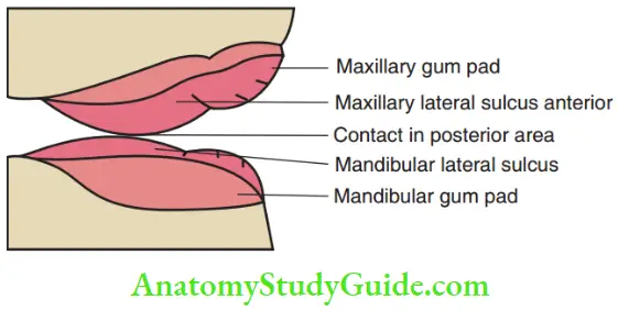
These gum pads house the developing dentition within them. The maxillary gum pad is oversized in both anteroposterior and transverse dimensions when compared with the mandibular arch of the gum pad. It appears as a bigger arch hovering over the mandibular arch with contact only at the molar region.
Lack of contact in the anterior region resembles an open bite and is called an infantile open bite. The presence of this open bite seems designed to accommodate the nipple and tongue to enable suckling action. The tongue is placed in the anterior open bite region, giving the appearance of an infantile tongue thrust during infantile swallowing.
On close observation, certain distinct morphological features are noticed on the gum pads:
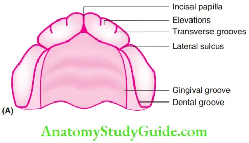
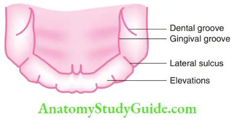
- Dental groove: It is a groove running along the length of the gum pads, dividing it into lingual or palatal and buccal or facial portions.
- Gingival groove: It is a groove running along and lingual or palatal to the dental groove.
- Elevations: Ten mild elevations are seen along the arch on each gum pad, divided by the transverse groove. Each elevation corresponds to a primary tooth developing beneath it.
- Transverse groove: It is transversely oriented and seen between elevations.
- Lateral sulcus: It is the most prominent and readily visible transverse groove found in between the canine and the fist molar area. The maxillary lateral sulcus is generally anterior to the mandibular lateral sulcus. This indication helps to predict interarch anteroposterior relationship of the maxilla and the mandible. Although it is a primitive prediction, its importance cannot be ruled out.
- Incisive papilla: Incisive papilla is seen in the maxillary gum pad in the midline between the anticipated places of the eruption of the right and left maxillary central incisors.
Deciduous Dentition Stage
Eruption of deciduous/primary teeth around 6 months of age marks the beginning of this stage. The eruption process of primary teeth lasts for 2.5–3 years. It extends from the eruption of the central incisors at around 6 months of age to the eruption of the second primary molars at around 2.5–3 years of age. Thus, 20 teeth are established in each arch.
The characteristics of this stage are as follows:
- Establishment of overbite and overjet by the eruption of the primary incisors in the region of the anterior open bite, eliminating the same
- Establishment of interdigitation (occlusion) by the primary molars guided by incisal stops
- Features The deciduous dentition is characterised by six features that are not considered ideal for permanent dentition. Some of these features aid in accommodating the erupting permanent teeth. The other features are settled during the transition of dentition. The features of the deciduous dentition are shown in Figure and discussed in detail next.
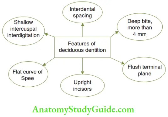
- Features The deciduous dentition is characterised by six features that are not considered ideal for permanent dentition. Some of these features aid in accommodating the erupting permanent teeth. The other features are settled during the transition of dentition. The features of the deciduous dentition are shown in Figure and discussed in detail next.
- Interdental Spacing Interdental spacing is a mild generalised or localised spacing between the primary teeth. It can be of two types, namely primary space and primate space.
- Primary Space The mild generalised spacing present between the deciduous anterior teeth is called primary spacing. Earlier terms such as developmental spacing and physiological spacing have become less relevant as these spaces do not develop or increase after the eruption of primary teeth. Primary spaces are observed in between the primary anterior teeth and amount to about 4 mm in the maxillary arch and 3 mm in the mandibular arch. A primary dentition with this space is called a spaced dentition. Primary spacing is useful to accommodate the larger-sized permanent incisors during the transition phase. They compensate for the incisal liability (explained later). Hence, a spaced deciduous dentition is likely to result in a well-aligned anterior segment of permanent dentition. When these spaces are absent, the dentition is termed non-spaced or closed dentition. A closed or non-spaced deciduous dentition is likely to result in a crowded anterior segment of permanent dentition unless compensated by spaces generated during growth.
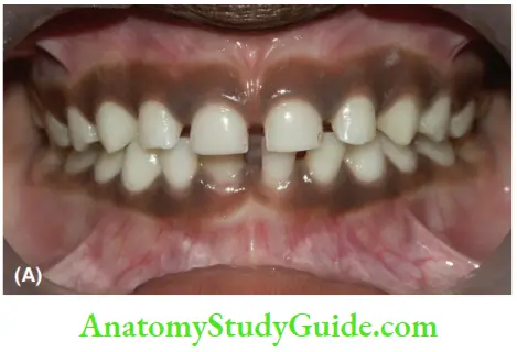
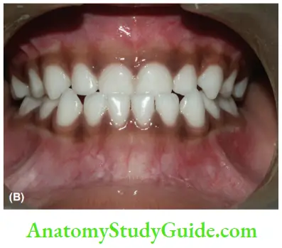
- Primate Space While mild generalised spacing of the anterior teeth is a common feature in the deciduous dentition, some children have a more prominent space in regions mesial to the maxillary deciduous canine and distal to the mandibular deciduous canine. These are called primate spaces. Since such space is a regular feature in primates, they are termed primate/simian/ anthropoid spaces. The maxillary and mandibular deciduous canines occlude in the corresponding primate spaces of the opposing arch. Primate spaces provide space to accommodate the larger permanent incisors, compensate for incisal liability (explained later) and contribute to the development of a well-aligned anterior segment. Primate spaces also allow an early mesial shift of molar to establish the appropriate molar relation.
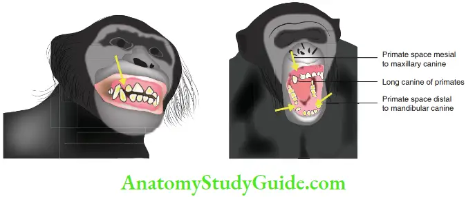
1. Primates have long canines and spaces between anterior teeth, mesial to the maxillary canine and distal to the mandibular canine, to accommodate the long canines of the opposing arch. Lack of such provision by nature might lead to constant injury to the lip and alveolus, decreased food intake and slow elimination of the species. These anthropoid spaces probably have helped the survival of species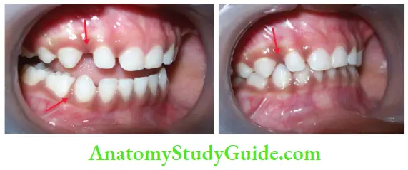
- Primary Space The mild generalised spacing present between the deciduous anterior teeth is called primary spacing. Earlier terms such as developmental spacing and physiological spacing have become less relevant as these spaces do not develop or increase after the eruption of primary teeth. Primary spaces are observed in between the primary anterior teeth and amount to about 4 mm in the maxillary arch and 3 mm in the mandibular arch. A primary dentition with this space is called a spaced dentition. Primary spacing is useful to accommodate the larger-sized permanent incisors during the transition phase. They compensate for the incisal liability (explained later). Hence, a spaced deciduous dentition is likely to result in a well-aligned anterior segment of permanent dentition. When these spaces are absent, the dentition is termed non-spaced or closed dentition. A closed or non-spaced deciduous dentition is likely to result in a crowded anterior segment of permanent dentition unless compensated by spaces generated during growth.
- Deep Bite Overbite is the overlapping of the lower anterior by the upper anterior. When the overlap is more than 4 mm, it is called a deep bite. A deep bite is considered as a malocclusion in permanent dentition and is acceptable in primary dentition. The eruption of permanent teeth and the dynamics of facial growth reduce the deep bite during the transition phase of dentition. Deep bite in primary dentition gradually gets reduced with further dynamic changes during the transition period, while a deep bite in permanent dentition initiates deterioration of the dentition. Spontaneously uncorrected deep bite also
has a restrictive effect on the expression of anterior mandibular growth.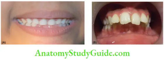
1. (A) Normal feature in deciduous dentition.2. (B) Malocclusion in permanent dentition with a deep bite.Deep bite gets reduced in permanent dentition by the following features:- Replacement of upright deciduous incisors with more proclaimed permanent incisors
- Attrition of lower deciduous incisors due to incisal wear
- Eruption of the permanent molars
- Downward and forward growth of the mandible in relation to the maxilla, carrying lower teeth along with it to a more downward and forward position
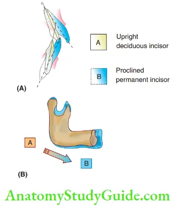
1. (A) Upright deciduous incisors cause the deep bite. More proclamation of the permanent incisors reduces deep bite.
2. (B) Downward and forward growth of the mandible from position A to B carries the lower teeth along and contributes to the reduction of deep bite.
- Flush Terminal Plane Flush means ‘on even level or same level’. The terminal plane is the end plane or the distal-most, true, perpendicular plane to the distal contour of the maxillary or mandibular second deciduous molars on a true lateral view. In more than 50% of the population, the maxillary and mandibular second deciduous molars occlude with their terminal planes at the same spatial plane (in a flash manner) and hence are said to be in an flsh terminal plane (FTP)/straight terminal plane relationship. In the rest of the population, the terminal planes exhibit two varied relationships, namely a mesial step and a distal step relationship.
- The significance of FTP is explained: Significance of the Flush Terminal Plane (FTP)
- Primary mandibular molars are wider mesiodistally than their maxillary counterparts. A longer arch length is left when they exfoliate. This difference in arch length leads to a more mesial movement of mandibular first permanent molars compared with maxillary molars during transition and settling of occlusion. The differential mesial movement of upper and lower molars enables them to settle in an ideal Class I molar relationship only when they begin their journey from an FTP status. An FTP relationship is therefore a favourable condition.
- The distal and mesial steps of the FTP affect the position of molars in full occlusion. The distal step and mesial step are likely to result in a Class II and Class III molar relationship, respectively. Molars in a non-FTP relationship depend on other factors such as spacing in the arch, vector of growth, cephalocaudal growth gradient and functional forces for achieving an ideal occlusion. When the arch perimeter is intact with no loss of tooth material, a distal step or mesial step of the FTP is an indication of a likely anteroposterior discrepancy of the dentoalveolar or maxillomandibular skeletal relationship. The distal step or mesial step of the FTP requires an assessment of leeway spaces and skeletal growth trends. Identification, localisation of the problem, timely intervention and early guidance at this stage are essential to achieve an ideal future occlusion.
- When the terminal plane of the mandibular deciduous second molar lies mesial to its maxillary counterpart, it is said to be in a mesial step relationship. The terminal plane of the mandibular deciduous second molar lies distal to its maxillary counterpart, it is said to be in a distal step relationship (discussed under section Mixed Dentition Period).
- The significance of FTP is explained: Significance of the Flush Terminal Plane (FTP)
- Upright Incisors Primary incisors erupt in an upright position without much labial inclination. The arch perimeter seems adequate in most conditions except in cases of crowded primary dentition. There is inadequate space when larger permanent incisors attempt to erupt in the same arch space occupied by smaller primary incisors. This liability status is managed by an increased labial inclination of the permanent incisors.
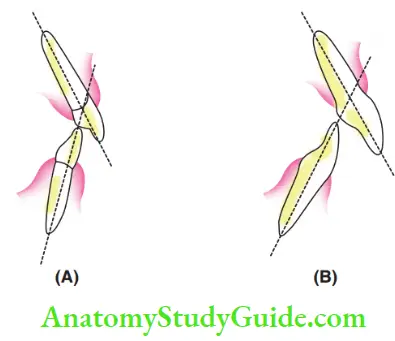 1. (A) Upright deciduous incisors.
2. (B) Increased labial inclination of permanent incisors. - Flat Curve Of Spee The curve of Spee is defined as the curvature of the mandibular occlusal plane. It begins at the tip of the lower canine and follows the buccal cusps of the posterior teeth, continuing up to the terminal molar. The acceptable depth of the curve of Spee in permanent dentition is about 2–4 mm. In primary dentition, the curve is almost flat, in the absence of premolars.
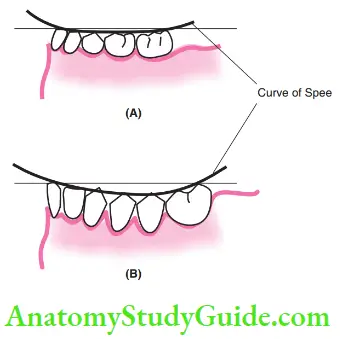
1. (A) when compared with that in permanent dentition (B) - Shallow Intercuspal Interdigitation The anatomical cuspal pattern is well-defined with proper cuspal elevations and deep occlusal fossae in permanent dentition. The morphology of cusps and grooves is less defied in the primary dentition. Hence, the interdigitations are also shallow in the primary dentition.
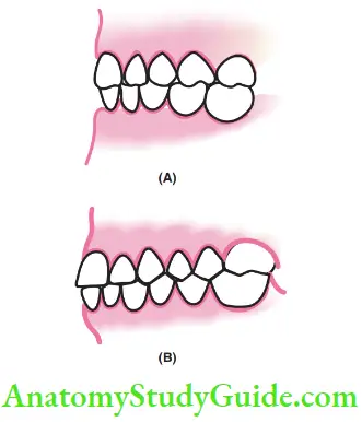
- Clinical Consideration As deciduous teeth form the basic infrastructural template and guide the eruption and occlusion of permanent dentition, it is essential to preserve their integrity and teeth material. This can be done by caries prevention and appropriate restorations. Extractions of deciduous teeth should be deferred and pulpal therapies and restoration of deciduous teeth should be sought as deciduous teeth are the best space maintainers.
Mixed Dentition Stage
The mixed dentition period is the transition phase from deciduous dentition to permanent dentition where both deciduous and permanent teeth are present in the arch. This stage lasts for a period from 7–8 years of age to 12–13 years of age. The eruption of a permanent tooth marks the beginning of the mixed dentition phase.
The eruption of the last succedaneous permanent tooth marks the end of this phase. At this stage, the developing permanent teeth within the alveolar process start migrating towards the alveolar crest, while apices of the corresponding deciduous teeth get resorbed.
Each deciduous tooth guides its successor along the path of eruption. The transition phase can be better described or understood when categorised into the first, inter and second transitional phases.
- First Transition Phase The two significant events at this phase are the eruption of the first permanent molar and the exchange of incisors.
- Eruption of the First Permanent Molar Mandibular first permanent molars followed by maxillary first permanent molars are usually the first permanent teeth to erupt in the oral cavity at around 6yearsofage. Hence, they recalled the sixth-year molars. The eruption of the first permanent molars is guided by the distal surfaces of roots and the terminal planes of the second deciduous molars. Settlement of occlusion and interdigitation of the opposing first permanent molars depends on the following three factors:
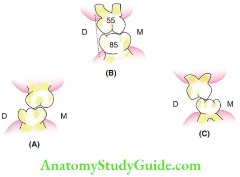
- Terminal plane status: As mentioned earlier, an FTP is likely to result in Class 1 molar relationship. Distal step and mesial step terminal planes are likely to guide permanent molars to a Class 2 and a Class 3 relationship, respectively. The position of the erupting permanent molars and the corresponding molar relationships are depicted in Figure and discussed later in this chapter.
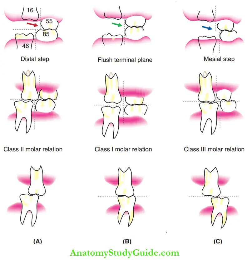 1. (A) Distal step of the terminal plane (red arrow) guiding the permanent molar to Class 2 relationship.
2. (B) Flush terminal plane (green arrow) guiding the permanent molar to cusp to the cusp, end-on and later Class 1 relationship.
3. (C) Mesial step of the terminal plane (blue arrow) guiding permanent molar to Class 3 relationship. (55, deciduous maxillary second molar; 85, deciduous mandibular second molar; 16, permanent maxillary first molar; 46, permanent mandibular first molar.) - Presence/absence of spaces: In a spaced dentition, the erupting mandibular first permanent molars exert a mesial force on the primary molars. The primary molars drif mesially and this is termed an early mesial shift With an early mesial shift the FTP is converted into a mesial step. Consequently, the permanent molars erupt into a Class 1 relation. In a non-spaced dentition, an early mesial shift does not occur and the permanent molars erupt into an end-on relationship.
- Cephalocaudal growth gradient: During postnatal growth, all the parts of the body do not grow in proportion. The parts away from the head end or the cephalic end grow at a greater proportion than the ones near the head end. There is a gradual increase in the proportion of growth as the position moves caudally. This phenomenon is called the cephalocaudal growth gradient. Due to this growth gradient, the magnitude of growth this relatively more in the mandible than in the maxilla. Therefore, the mandibular teeth are carried more forward than the maxillary teeth converting an FTP into a mesial step. The mesial step guides the first permanent molars into a Class I relation. To conclude, the terminal plane status, presence/absence of spaces in deciduous dentition and the magnitude of the cephalocaudal growth gradient act in a composite manner to determine the permanent molar relationship. This warrants periodic monitoring to initiate preventive/interceptive orthodontic treatment strategies.
- Terminal plane status: As mentioned earlier, an FTP is likely to result in Class 1 molar relationship. Distal step and mesial step terminal planes are likely to guide permanent molars to a Class 2 and a Class 3 relationship, respectively. The position of the erupting permanent molars and the corresponding molar relationships are depicted in Figure and discussed later in this chapter.
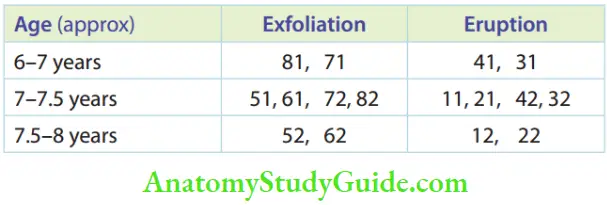
- Exchange of Incisors The exfoliation of primary incisors and subsequent eruption of permanent incisors begin at around 7 years of age. Usually, it is the lower central incisors that start the cascade of events. Permanent lower incisors erupting lingual to deciduous incisors are moved into the arch by tongue pressure. A pre-eruptive bulge around a primary incisor precedes the eruption of the permanent incisor. The combined mesiodistal width of permanent incisors is distinctly larger than the combined mesiodistal width of deciduous incisors. Hence, accommodating larger teeth in the smaller space available is a liability or a burden. This was termed incisal liability by Warren Mayne in 1969. The incisal liability can be equated.
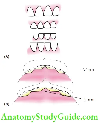
1. (A) Larger permanent incisors are waiting to erupt into a place occupied by smaller deciduous incisors.
2. (B) If the deciduous incisors occupy x mm and the permanent incisors need y mm, x – y is the incisal liability.- Incisal Liability
- Space equation for the anterior segment of the maxillary arch
- Space available = Mesiodistal width of primary incisors = x mm
- Space required = Mesiodistal width of permanent incisors = y mm
- Space discrepancy/shortage = x – y = –7.6 mm approximately
- Space equation for the anterior segment of the mandibular arch
- Space available = Mesiodistal width of primary incisors = x1 mm
- Space required = Mesiodistal width of permanent incisors = y1 mm
- Space discrepancy/shortage = x1 – y1 = –6 mm approximately
- The incisal liability of 7.6 mm in the maxillary arch and 6 mm in the mandibular arch can appropriately be nullified if the following three factors donate adequate space:
- Interdental spaces in the primary dentition (2–3 mm): Primary and primate spaces alleviate the incisal discrepancy. Non-spaced or closed dentition or a crowded deciduous dentition is likely to cause crowded permanent incisors. Therefore, when no natural spacing exists, the situation is solely dependent upon the next two factors to overcome the incisal liability.
- Increase in inter canine width (3–4 mm): Growth is expressed in all three dimensions contributing to an increase in arch length, arch width and arch perimeter. An increase in inter canine width provides a 3–4 mm increase in arch perimeter to accommodate the four large permanent incisors.
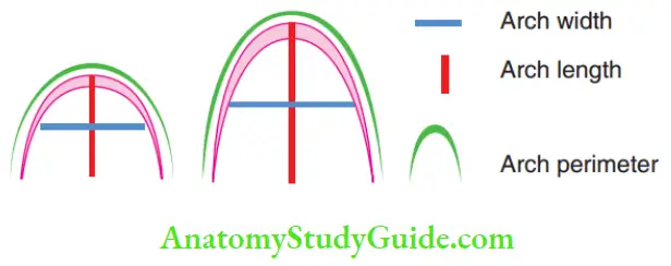
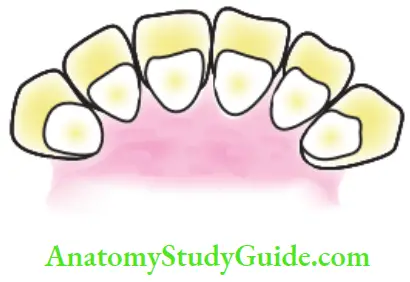
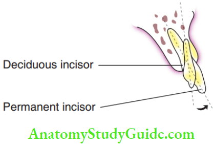
- Change of inclination and positioning of permanent incisors (1–2 mm): Th axial inclination of the permanent incisors is more labial than that of the primary incisors. The permanent incisors are placed more labially than the primary teeth. This change in angulation (axial inclination) and labial positioning of the permanent incisors make the dental arch larger providing 1–2 mm to reduce the incisal liability.
- The incisal liability of 7.6 mm in the maxillary arch and 6 mm in the mandibular arch can appropriately be nullified if the following three factors donate adequate space:
- Incisal Liability
- Eruption of the First Permanent Molar Mandibular first permanent molars followed by maxillary first permanent molars are usually the first permanent teeth to erupt in the oral cavity at around 6yearsofage. Hence, they recalled the sixth-year molars. The eruption of the first permanent molars is guided by the distal surfaces of roots and the terminal planes of the second deciduous molars. Settlement of occlusion and interdigitation of the opposing first permanent molars depends on the following three factors:
- Intertransitional Phase This phase is apparently a nil activity phase between the two active transitional periods, lasting for about 1–1.5 years. There is no exchange of teeth or no significant events. The functional forces exerted by soft tissues during speech, swallowing, etc., play a great role in settling the position of erupted teeth. Lip and tongue pressure play an important role in deciding the final position of permanent incisors. Growing jaws carry the incisors to a downward and forward position and thrust them into the lips. The lip pressure restricts further forward proclamation of the incisors. This, the incisors are moulded to their position between the tongue and the lips in the growing jaw. showing the teeth that are present and their position within the alveolus and bone in the enter transition period.
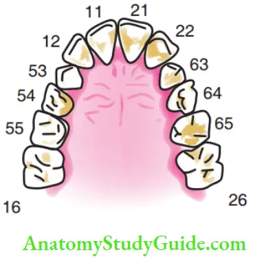
1. The permanent incisors (11, 21, 12, 22), first permanent molars (16, 26), deciduous canines (53, 63) and deciduous molars (54, 64, 55, 65)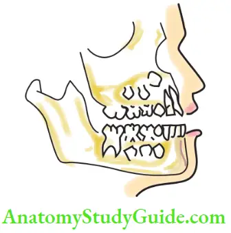
- Second Transitional Phase This phase demonstrates the exchange of canines and primary molars and establishes proper selling of posterior occlusion. The exfoliation sequence of primary canines and primary molars and the eruption sequence of permanent canines and premolars are described. The tooth buds of the permanent maxillary canine are near the infraorbital margin and travel the longest course than any other tooth during eruption. The maxillary canine seeks its way between the lateral incisor and the fist premolar, which have already taken their positions in the arch. The canines make way between the roots of these two teeth, pressurising them to move away from the path.
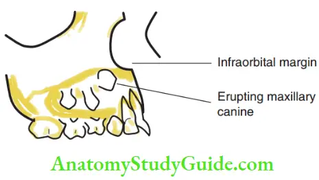
- Ugly Duckling Stage At the time of the eruption of the canine, an interesting transient malocclusion called the ugly duckling stage or Broadbent phenomenon occurs in the maxillary arch. The following events occur during the process of eruption of the maxillary canine. Maxillary canine descends from the region of the infraorbital margin to the root level of the lateral incisor.
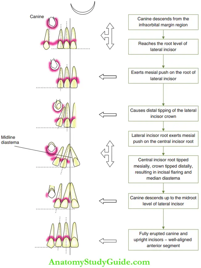
- The descending canine exerts a mesially directed push upon the root of the lateral incisor due to which the crown of the lateral incisor flies with a distal tip.
- The lateral incisor root, in turn, transmits a mesially directed push on the root of the central incisor.
- The central incisor flies with the root tipped mesially and crown tipped distally, resulting in a ‘V’-shaped median diastema.
- Self-correction of the ugly duckling stage begins to occur when the canine descends up to the mid-root level of the lateral incisor.
- The mesially exerted pressure on the lateral incisor disappears and this allows the incisors to be upright.
- The median diastema begins to close and disappears when the canine reaches the occlusal plane.
- Any attempt to correct the diastema should be deferred during this stage.
- The strikingly altered appearance of the child perturbs parents and they seek treatment for the transient malocclusion. the figure shows three cases.
- shows a child with intact primary dentition that is acceptable aesthetically.
- shows clinical views and the radiograph of mixed dentition during the ugly duckling stage, where aesthetics is affected.
- The self-correction of this phenomenon takes a few months and is best left uninterrupted, yet monitored. Any attempt in closing the diastema with springs or other orthodontic means or correcting the axial inclination of the central and lateral incisors is most likely to result in root resorption of incisors, the altered path of eruption of canines or impaction of canines. A clinician has to clearly understand the above phenomenon, give assurance to the perturbed child and parents and periodically follow up the case explaining the transient nature of this self-correcting phase. The ugly duckling stage does not manifest in the mandibular arch, because two reasons:
- Mandibular canines erupt before the fist premolars and have a broader pathway without a need to exert force upon the neighbouring roots.
- Mandibular teeth do not have a long eruption path.
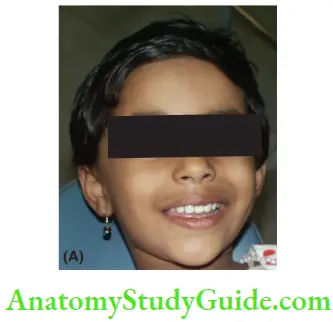
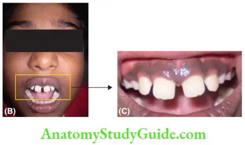
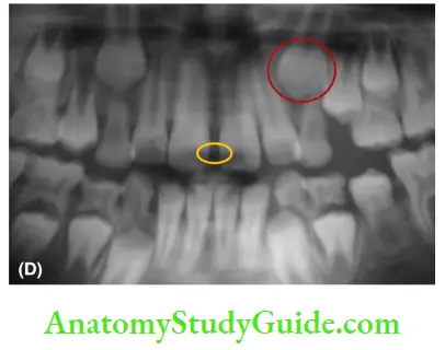
- The strikingly altered appearance of the child perturbs parents and they seek treatment for the transient malocclusion. the figure shows three cases.
- Ugly Duckling Stage At the time of the eruption of the canine, an interesting transient malocclusion called the ugly duckling stage or Broadbent phenomenon occurs in the maxillary arch. The following events occur during the process of eruption of the maxillary canine. Maxillary canine descends from the region of the infraorbital margin to the root level of the lateral incisor.
- Leeway Space of Nance Contrary to the inadequacy of space for the erupting permanent incisors, there usually exists an excess of space for the erupting permanent canines and premolars, as the smaller-sized premolars replace larger-sized primary molars. The combined mesiodistal width of the deciduous canine and primary molars is greater than the combined mesiodistal width of the permanent canine and premolars. This discrepancy of excess space available is termed the leeway space of Nance. The calculation is shown.
- Calculation of Leeway Space
- Space equation in the posterior segment of the maxillary arch (per side)
- Space available: Mesiodistal width of primary canine and molars = R mm
- Space required: Mesiodistal width of permanent canine and premolars = S mm
- Space excess = R – S = 0.9 mm approximately
- Space equation in the posterior segment of the mandibular arch (per side)
- Space available: Mesiodistal width of primary canine and molars = T mm
- Space required: Mesiodistal width of permanent canine and premolars = U mm
- Space excess = T – U = 1.7 mm approximately
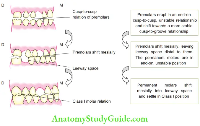
- The leeway space of Nance amounts to 1.7 mm per side and cumulates to 3.4 mm in the mandibular arch. The leeway space of the maxillary arch amounts to 0.9 mm per side cumulating to 1.8 mm. The calculated leeway space value is an average and the exact leeway space for an individual has to be calculated using the measurement of the individual’s teeth. The Leeway space of Nance is utilised in the second transitional period by mesial migration of the permanent molars. This is termed the late mesial shift and it establishes a permanent molar relationship and decreases the arch length. The leeway space is higher in the mandibular arch. This transforms an FTP, primary molar relation (Rendon permanent molar) into a Class 1 permanent molar relation. The availability of leeway space is essential for the proper settling of the cusp-to-groove relationship of the posterior teeth. When leeway space is lost due to the premature loss of deciduous posterior teeth, the neighbouring teeth migrate into the space and eliminate its advantage. The significant activities during all three phases of the mixed dentition stage are depicted in Figure.
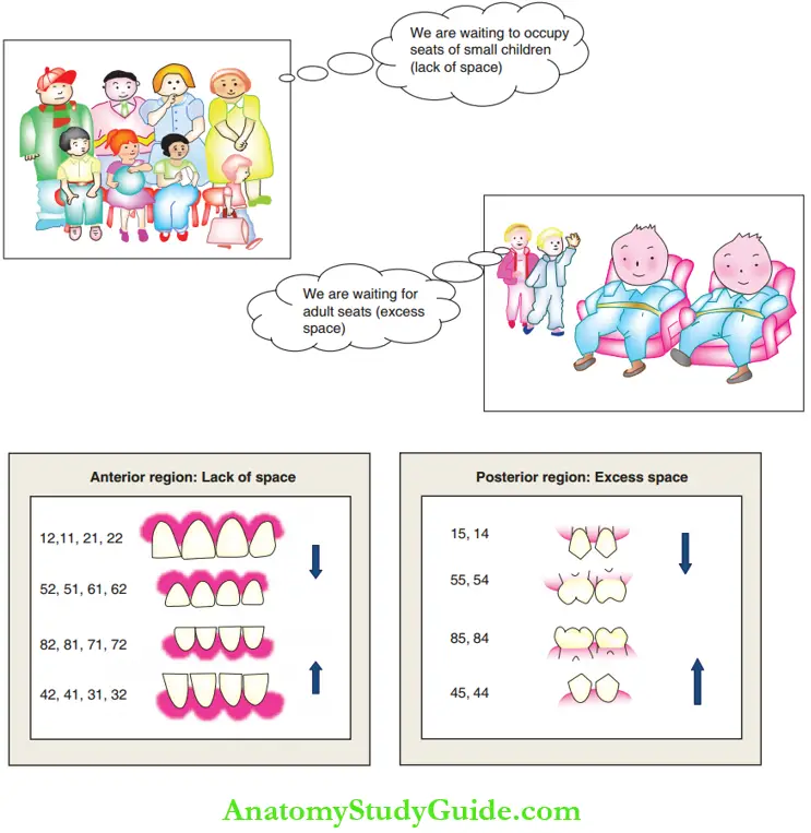
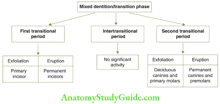
- The leeway space of Nance amounts to 1.7 mm per side and cumulates to 3.4 mm in the mandibular arch. The leeway space of the maxillary arch amounts to 0.9 mm per side cumulating to 1.8 mm. The calculated leeway space value is an average and the exact leeway space for an individual has to be calculated using the measurement of the individual’s teeth. The Leeway space of Nance is utilised in the second transitional period by mesial migration of the permanent molars. This is termed the late mesial shift and it establishes a permanent molar relationship and decreases the arch length. The leeway space is higher in the mandibular arch. This transforms an FTP, primary molar relation (Rendon permanent molar) into a Class 1 permanent molar relation. The availability of leeway space is essential for the proper settling of the cusp-to-groove relationship of the posterior teeth. When leeway space is lost due to the premature loss of deciduous posterior teeth, the neighbouring teeth migrate into the space and eliminate its advantage. The significant activities during all three phases of the mixed dentition stage are depicted in Figure.
- Clinical Considerations
- Calculation of Leeway Space
- Over-Retained Deciduous Tooth Any deciduous tooth present in the arch beyond its expected range of exfoliation time is termed an over-retained tooth. Deciduous teeth are over-retained due to either the congenital absence of a succedaneous tooth or an altered eruptive path of the successor. Certain systemic diseases such as hypothyroidism also cause over-retention of teeth.
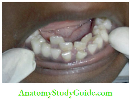
- Submerged Molar An ankylosed over-retained deciduous second molar is called a submerged molar. When the permanent second premolar is congenitally absent, its predecessor, the second deciduous molar, is over-retained. With a shorter crown height, the over-retained molar seems submerged when compared with its neighbours, the first premolar and first permanent molar, thus earning a name, submerged molar. A submerged molar does not contribute to leeway space and is likely to jeopardise molar occlusion.

Permanent Dentition Stage
Permanent dentition is established by around 12–13 years of age when all the deciduous teeth are replaced by their permanent successors. The second and third permanent molars erupt subsequently. Eruption of the second permanent molar at around 14 years of age contributes to the second phase of bite opening. The third permanent molar shows the most variability in its size, shape and position and eruption time. It erupts by around 16 years of age and contributes to the third phase of bite opening. Impacted third molars are witnessed frequently at varying levels and positions. The permanent maxillary and mandibular dentition generally settle in the following three types of relationship with each other:
- Disto-occlusion or Class 2 condition
- Neutro-occlusion or Class 1 condition
- Mesial-occlusion or Class 3 condition
- In most individuals, the Class 1, Class 2 and Class 3 dental patterns correlate with their Class 1, Class 2 and Class 3 types of skeletal jaw base relationship, respectively. The facial profile is an indicator of the skeletal jaw-base relationship. The facial profile posteriorly diverges in Class 2, is straight in Class 1 and anteriorly diverges in Class 3 skeletal conditions. Settlement of occlusion in the permanent dentition is affected by hereditary and environmental factors. The presence of the ideal number, size, shape and consistency of teeth with intact tooth material is essential for ideal occlusion. Teeth size corresponding to arch size is another deciding factor. Systemic conditions also affect occlusal relationships.
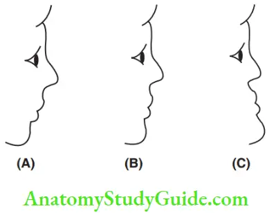 1. (A) Class 2 (posteriorly diverging),
2. (B) Class 1 (straight) and (C) Class 3 (anteriorly diverging)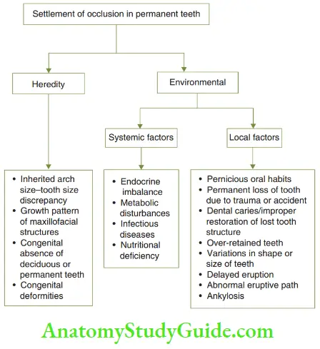
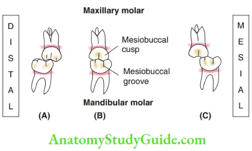
- Angle’S Classification Molars, canines and incisors are described in terms of Class 1 or Class 2 or Class 3 relationship with their opposing counterparts. The molar relationship alone is called Angle’s classification after Edward Angle. Angle’s Class 1 molar relationship is where the mesiobuccal cusp of the maxillary first permanent molar occludes with the mesiobuccal groove of the mandibular first permanent molar. When the mesiobuccal cusp of the maxillary first permanent molar occludes in a more distal or mesial relationship with the mandibular first permanent molar, the condition is described as Class 2 and Class 3 molar relationship, respectively. When the maxillary molar cusps occlude upon the corresponding mandibular molar cusps, it is described as an end-on relationship.
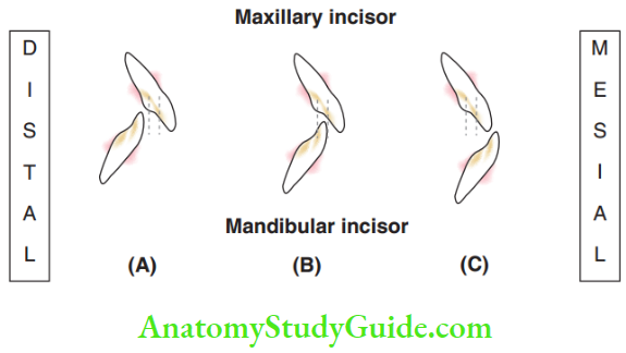
- Incisor And Canine Relationship When the lower incisor’s incisal edges lie within the region of the cingulum plateau of the upper incisors and the central axis line of a normally placed upper canine lies tangential to the distal surface of the lower canine, it is described to be in a Class I relationship of incisors and canines, respectively. Similar to the molars, when the lower incisors and canines are positioned in a more distal or mesial relationship to their upper counterparts, they are described to be in Class II and Class III relationship,
respectively.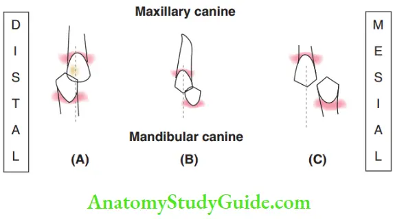
Keys Of Occlusion
Andrews described six requisites for the ideal occlusion of permanent dentition, known as the six keys of occlusion. They are as follows:
- Key 1: Molar relationship: Mesiobuccal cusp of the fist maxillary permanent molar rests on the mesiobuccal groove of the fist mandibular permanent molar.
- Key 2: Crown angulation (tip): Gingival aspects of teeth are mildly distal to the incisal or occlusal aspect.
- Key 3: Crown inclination (torque): Th gingival portion of the anterior teeth is more lingual than their incisal ends, with a mild positive crown torque. The posterior crowns are lingually inclined with a mild negative torque.
- Key 4: Absence of rotation of any tooth: Rotations of the anterior teeth occupy less space and those of the posterior teeth occupy more space in the arch. The absence of rotation is important for an ideal occlusal relationship.
- Key 5: Interdental contacts: All the teeth should be in close proximity with tight contact without interdental spacing for ideal occlusion.
- Key 6: Curve of Spee: Th occlusal plane is flat or mildly curved; the curve of Spee not exceeding 1.5 mm. Bolton suggested a tooth ratio as an additional or seventh key to occlusion:
- Bolton’s seventh key of occlusion: Bolton’s tooth ratio states that an ideal ratio between the maxillary and mandibular tooth material gives rise to an ideal interdigitation.
Transient Malocclusion
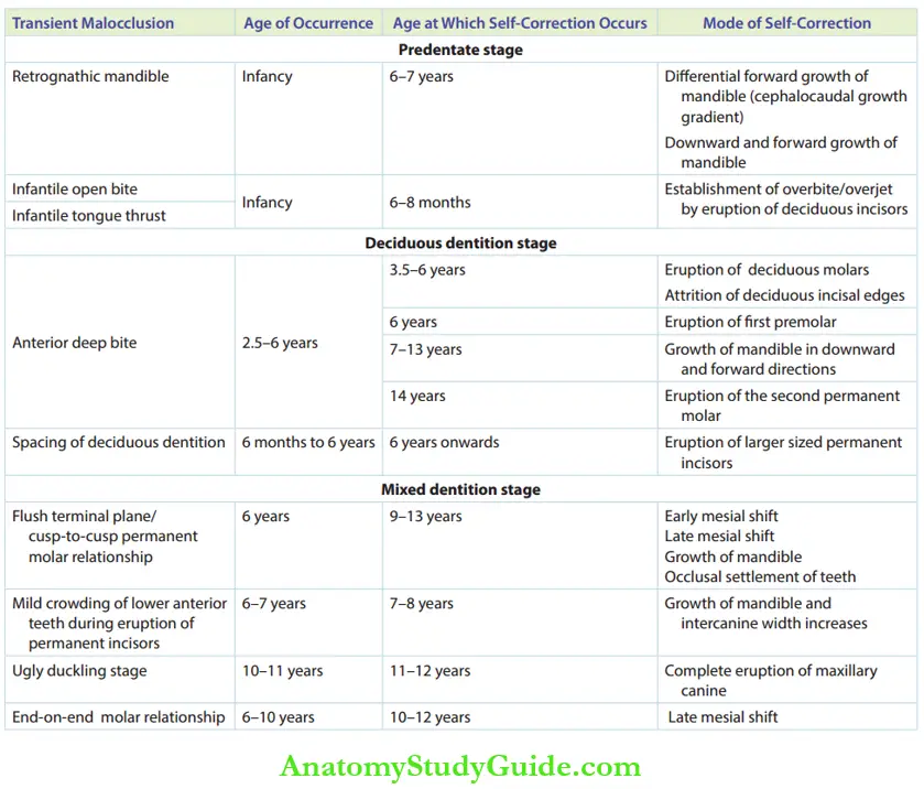
- Transient malocclusions are temporary irregularities of teeth positions or discrepancies in jaw relationship. These conditions are self-correcting with normal growth and development and do not require active intervention.
- Attempts to correct them with active therapy will initiate iatrogenic malocclusion. A clinician should possess the knowledge to clearly identify and differentiate a transient malocclusion from an actual malocclusion requiring active intervention.
- The table summarises transient malocclusion conditions occurring in various stages of the development of dentition.
- Transient malocclusions require periodic reviews and observations only. The condition does not call for action unless the self-correction process seems to fail or delay. The child and parents have to be explained about the transient nature of the unaesthetic appearance and refrained from attempting intervention. Records like study models, photographs and selected radiographs will help in monitoring the progress of the situation.
Development Of Occlusion Summary
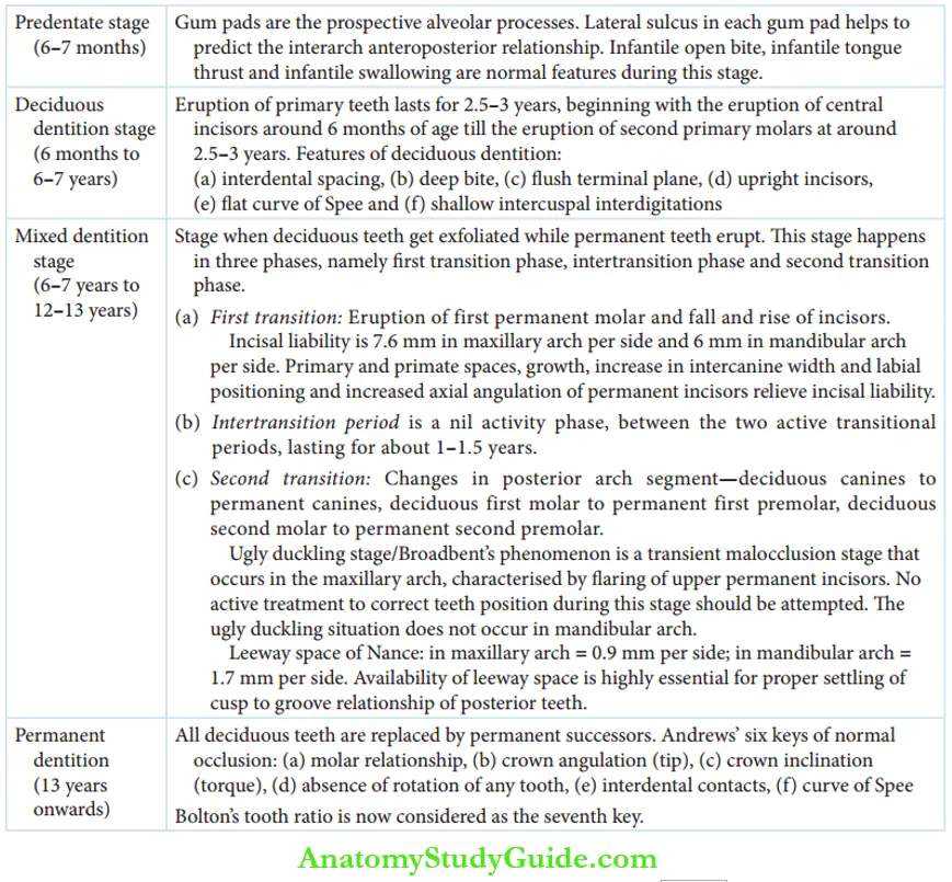
Development of occlusion can be distinctly identified as happening in four stages: Transient malocclusions/self-correcting malocclusions need periodic review and no active intervention
Terms
- Preprimary teeth or natal teeth: Teeth that have erupted into the oral cavity at birth in an infant are called natal teeth. They neither have roots nor are fully attached to the alveolar ridge.
- Neonatal teeth: Teeth that erupt prematurely in the first 30 days of life are neonatal teeth.
- Primary spacing of deciduous dentition: Th mild generalised spacing in the anterior region of deciduous dentition.
- Primate/anthropoid/simian space: The most noticeable space mesial to the maxillary deciduous canine and distal to the mandibular deciduous canine resembling such spaces in primates.
- Flush/straight terminal plane: The terminal planes of maxillary and mandibular second deciduous molars lying in flesh or at the same level.
- Overjet: Th horizontal overlap of maxillary incisors over mandibular incisors is overjet.
- Overbite: Th vertical overlap of maxillary incisors over mandibular incisors is an overbite.
- Incisal liability: It is the difference between the amount of combined mesiodistal width deciduous incisors and that of permanent incisors. It is about 7.6 mm in the maxillary arch and 6 mm in the mandibular arch (right and left.
- Leeway space: It is the difference between the combined width of the permanent canine and premolars and that of the deciduous canine and molars. The surplus space is called leeway space. It is 1.8 mm in the maxillary arch and 3.4 mm in the mandibular arch (right and left.
- Ugly duckling stage: A transient malocclusion phase seen in the maxillary anterior region during the eruption of maxillary canines is called the ugly duckling stage. It is characterised by distal flying of permanent maxillary central and lateral incisors along with median diastema.
- Iatrogenic malocclusion: Treatment-induced malocclusion.
- Over-retained tooth: Deciduous tooth retained in arch beyond its normal range of exfoliation period.
- Submerged molar: The over-retained second deciduous molar amidst the first permanent molar and first premolar.
- Angle’s Class I molar relationship: The relationship between permanent fist molars when the mesiobuccal cusp of the maxillary first permanent molar occludes with the mesiobuccal groove of the mandibular first permanent molar.
- Transient malocclusion/self-correcting anomaly: These are malocclusion-like conditions existing for a brief period of time and self-rectifying in due course.
Leave a Reply