Embryonic Disc
Notochord (Noto—relationship to back, chord—rod-shaped structure)
Introduction: It is a solid rod of cells that supports the embryo.
Table of Contents
1. Extent: It extends from the primitive node to the prochordal plate.
2. Situation: Midline of the embryo.
3. Source: Cells of the primitive node.
Read And Learn More: General Histology Question And Answers
4. Formation:
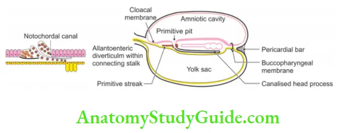
1. Cranial: The cranial end of primitive streak (streak—faint, white trace) becomes thick. This thickened part of primitive streak is called primitive node (knot or Hensen’s node).
- Depression appears: A depression appears: in the centre of primitive knot—called blastopore.
- Notochordal process: The cells of primitive node present in the central axis migrate cranially towards prochordal plate.
- Notochordal canal: The canal that arises from the blastopore and extends into the notochord is called the notochordal canal.
- Neuroenteric canal: The cells at floor of the notochordal canal break and form a communication between the amnion and yolk sac through the canal.
- Notochordal plate: The cells in the floor of the neurenteric canal reappear. They fill a gap at the floor and form a plate. Such a plate is called a notochordal plate
- Solid notochord: The proliferation of cells of the notochordal plate gives rise to solid notochord, which extends from the prochordal plate to primitive node.
5. Functions:
- It supports the embryo.
- It acts as a vertebral column in embryonic life.
- It induces surface ectoderm to form a neural plate.
6. Fate: It does not have any contribution for formation of vertebrae. It remains as ANC
- Apical ligament of dens.
- Nucleus pulposus of the intervertebral disc.
- Coccygeal body
7. Applied anatomy: Following are the anomalies
- Neurogenetic canal: Notochord partly remains patent. It connects to intestinal lumen to central canal of spinal cord.
- Notochordal: Notochordal cells in body of vertebrae proliferate to form chordoma.
Connecting stalk
Introduction: It is a part of extra-embryonic mesoderm which is not encroached by extra-embryonic coelom.
1. Formation:
- As the embryo grows, the size of the stalk becomes relatively small.
- Gradually, the attachment is shifted to the caudal end of the embryonic disc.
- With the rotation of the tail fold, attachment of the stalk moves and gets attached to a region of the umbilical opening.
- The blood vessels develop in embryo and placenta.
- The blood vessels of the placenta communicate with the blood vessels of the mother.
2. Contents:
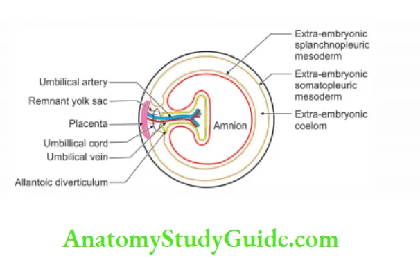
- Two umbilical arteries and one umbilical vein.
- The vitellointestinal duct may remain as remnants of the yolk sac.
- The mesoderm of connecting stalk forms a gelatinous substance called Wharton’s jelly.
3. Functions:
- It protects the umbilical vessels.
- It forms small part of the extra-embryonic coelom.
- It increases the length of the umbilical cord to allow free movement of the embryo.
- It suspends the embryo.
- It nourishes the embryo.
- It provides space for a physiological hernia.
4. Fate:
It gets converted into an umbilical cord
Umbilical cord
Introduction: It is a cord extending from the umbilicus of the fetus to the placenta.
1. Embryonic tissue:
After folding of embryo, connecting stalk elongates and forms the umbilical cord.
2. Dimensions:
- Length: 50 cm
- Diameter: 1-2 cm
3. Content:
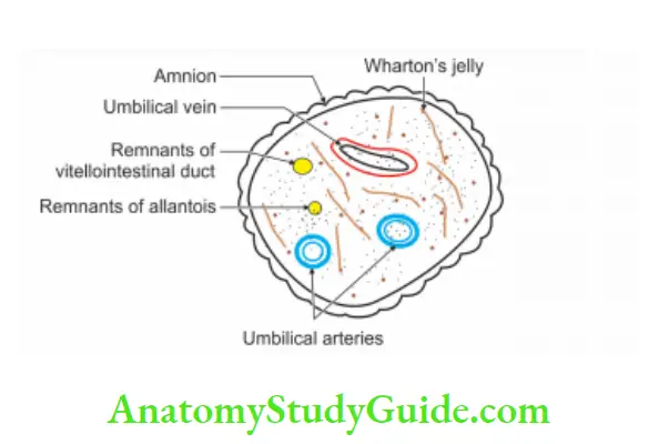
1. Vessels
- Two umbilical arteries.
- One umbilical vein.
2. Mesoderm: Intra-embryonic mesoderm called Wharton’s jelly.
3. Remnant structures: Vitellointestinal duct and allantois.
4. Layer of amnion.
4. Functions:
- It suspends fetus into amniotic cavity.
- It transfers nutrients.
5. Knots of umbilicus: These are
- True knots of umbilicus: These are due to excessive movements.
- False knots of umbilicus: These are due to sharp bend of the cord.
Intra-embryonic mesoderm (secondary mesoderm)
Introduction:
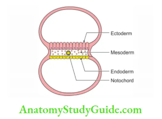
1. It forms the 3rd germinal layer.
2. It is present between ectoderm and endoderm .
1. Chronological age: It is formed in the 3rd week of gestation.
2. Sources: The cells of
- Primitive streak.
- Prochordal plate.
- Neural crest.
3. Derivatives of mesoderm:
1. Paraxial mesoderm
- Somites:
Sclerotome:
- Vertebrae,
- Portions of the neurocranium, and
- Axial skeleton.
Myotome: All voluntary muscles of the head, trunk and limb.
Dermatome: Dermis of skin over the dorsal region.
2. Neuromeres (head mesoderm): Endosteal layer of dura mater.
2. Intermediate cell mass or column:
- Connective tissue of the gonads.
- Mesonephric and metanephric nephrons.
- Smooth muscles and connective tissues of the reproductive system.
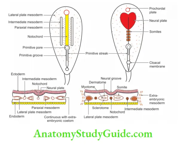
3. Lateral plate or column:
1. Septum transversum: It gives rise to
1. Structures related to heart:
- Epicardium
- Fibrous pericardium
2. Structures related to liver:
- Sinusoids of liver
- Falciform ligament
3. Central tendon of the diaphragm, and
4. Oesophageal mesentery.
2. Splanchnopleuric layer:
It gives rise to smooth muscle and connective tissues of the
- Intestinal tract and associated glands.
- Respiratory tract and associated glands.
3. Somatopleuric layer:
- Appendicular skeleton
- Connective tissue of limbs and trunks (It includes cartilage, ligament and tendons.),
- Dermis of the ventral part of the body and limbs, and
- Mesenchyme of external genitalia.
4. Angiogenic mesoderm:
- Endocardium of the heart.
- Endothelium of the blood and lymphatic vessels.
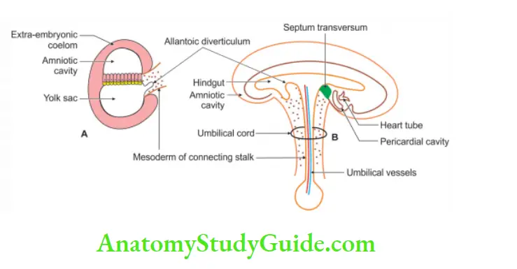
Allantois (allantois—sausage, eidos form)
Introduction: It is the diverticulum developing from the hindgut of the fetus.
1. Evolution:
- It is tubular ventral diverticulum in embryos of reptiles, birds and mammals.
- It is expanded to a large sac for storing urine in reptiles and birds.
- It is prominent in some mammals (carnivores and ungulates)
- It is vestigial in man.
2. Formation:
Before the formation of tail fold, a small endodermal diverticulum arises from the yolk sac near the caudal end of the embryonic disc.
This diverticulum grows into the mesoderm of the connecting stalk. After the formation of the tail fold, part of this endoderm is absorbed into the hindgut. It passes from the ventral side of the hindgut into the connecting stalk.
The developing bladder is continuous with the allantois. The allantois atrophies and is seen in postnatal life as a fibrous band termed as urachus. The urachus extends from the apex of the bladder to the umbilicus as median umbilical ligament
3. Anomalies:
1. Patent urachus: Urachus may remain patent and urine may pass through the umbilicus.
2. Urachal cyst: It results from partial persistence of the intra-embryonic allantois. The secretory activity of the linings of allantois produces the dilatation (cyst).
3. Urachal sinus: The lumen of the lower part of allantois persists. It causes the urachal sinus. The sinus is usually continuous with the urinary bladder.
4. Urachal fistula: An abnormal passage is formed from a patent urachus. It communicates with the umbilicus and urinary bladder
Septum transversum
1. Introduction: It is unsplit part of lateral plate mesoderm. It is present
cranial to the prochordal plate and intra-embryonic coelom.
2. Derivatives of septum transversum:
1. Structures related to heart:
- Epicardium
- Fibrous pericardium
2. Structures related to liver:
- Sinusoids of liver, and
- Falciform ligament.
- Central tendon of the diaphragm
- Oesophageal mesentery
Leave a Reply