Morphology Of The Pulp
Dental pulp is a soft, highly vascularized connective tissue present in the pulp chamber of the tooth. The dental pulp is enclosed by dentin on all the sides inside the pulp chamber. The pulp consists of nerves and blood vessels and various cells. It plays an important role in nutritive, defensive and sensory functions.
Table of Contents
A clear understanding of the pulp morphology is important for good clinical practice. Thorough knowledge about pulp morphology also plays an important role in treatment planning.
Read And Learn More: Oral Anatomy Notes
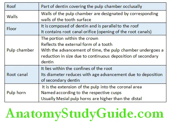
Terminologies Related To Pulp Morphology
Few important terminologies that need to be understood before progressing to individual tooth pulp morphology are given in table.
Terminology related to pulp morphology:
Root Canal Classification
Morphology of the root canal is highly complicated. Thus, numerous classifications have been introduced to classify root canals. The important ones are discussed as follows.
Weine’s classification:
Type 1: Single canal from pulp chamber to apex
Type 2: Two canals leaving the chamber and merging to form a single canal short of the apex
Type 3: Two separate and distinct canals from chamber to apex
Type 4: One canal leaving the chamber and dividing into two separate and distinct canals.
Pulp Morphology In Permanent Teeth
Maxillary central incisor:
Average length: 23.3 mm
Pulp chamber:
- Located in the centre of the crown
- Three pulp horns present
- Ovoid in shape mesiodistally
- Wider mesiodistally than buccolingually
Root and root canals:
- Single root with one root canal
- Root canal – broad labiolingually
Maxillary lateral incisor:
Average length: 22.5 mm
Pulp Chamber:
- Similar to central incisor but smaller pulp chamber.
- It has only two pulp horns.
- The shape of the pulp chamber may be round, oval or triangular.
- Pulp chamber is wider mesiodistally.
Root and root canals:
- Conical in shape; has an apical-distal curvature
- Usually one root canal but two or three canals have been reported
- Lateral canals are frequent (26%)
Maxillary canine:
Average length: 26 mm
Pulp chamber:
- Largest among any single-rooted teeth
- Wider labiolingually than mesiodistally
- It is triangular in shape labiolingually
- Flame shaped – mesiodistally
- Single pulp horn is present
Root and root canals:
- Single root
- Single canal is seen most often.
- Oval in shape, wider in labiopalatal direction
- The canal is straight in 39% of the cases
- Curved distally in 32% of the cases
Mandibular central incisor:
Average length: 20.8 mm.
Pulp chamber:
- Smallest in the arch
- Pulp chamber is flat mesiodistally, ovoid labiolingually
- It has three pulp horns when newly erupted, which calcify and disappear early because of constant masticatory stress
Root and root canals:
- One root with one canal (70%)
- One canal may bifurcate into two canals and exit via one apical opening (22%)
Mandibular lateral incisor:
Average length: 22.4 mm.
Pulp chamber:
- Same as the mandibular central incisor, but the lateral incisor has larger dimensions.
Root and root canals:
- Usually single root with single canal but two canals are also seen in few cases.
Mandibular canine:
Average length: 25 mm.
Pulp chamber:
- More wide labiolingually, ovoid, funnel-shaped
- Narrower mesiodistally
- Single pulp horn is present
Root and root canals:
- Single root
- Single canal (generally wide)
- Roots are usually straight (68%)
Maxillary first premolar:
Average length: 21.5 mm.
Pulp chamber:
- Narrow – mesiodistally
- Wide – buccopalatally
- One pulp horn under each cusp
- Buccal pulp horn is larger
- Wide and ovoid pulp chamber
- Convex pulpal floor of pulp chamber with two canal orifices
Roots and root canals:
- Usually has two roots and two canals (54.6%)
- Both separated and fused roots may be seen
- Three roots with three canals can also be seen in few of the cases
Maxillary second premolar:
Average length: 21.6 mm.
Pulp chamber:
- Similar to first premolar but narrower
- There are two pulp horns
- The roof is similar to the first maxillary premolar
Root and root canals:
- Usually single rooted (90.3%) with single canal but may have two or three canals.
- Single root is oval and wider.
Mandibular first premolar:
Average length: 22.1 mm.
Pulp chamber:
- It is narrow mesiodistally and wider buccolingually.
- It has a prominent buccal pulp horn.
- Pulp chamber is ovoid buccolingually.
Root and root canal:
- Single root with one canal (cone shaped)
- Root is mesiodistally narrow and buccolingually wider
Mandibular second premolar:
Average length: 22.3 mm.
Pulp chamber:
- Similar to first mandibular premolar
- Lingual horn more prominent
Roots and root canal:
- Usually single rooted with a single canal
Maxillary first molar:
Average length: 21.3 mm.
Pulp chamber: Largest in the dental arch
- Four pulp horns – mesiobuccal, distobuccal, mesiopalatal, distopalatal. Therefore, pulpal roof has a rhomboidal appearance
- Floor of the chamber is triangular in shape with apex at the palatal orifice
Root and root canals:
- Three roots – three canals: Mesiobuccal, mesiolingual and palatal
- Four canals may be seen (mb2 canal)
- Mesiobuccal root is narrower and smaller
- Usually mb roots have a distal curve
- The apical distal curvature of the mesial root is seen in 78% of teeth
- Palatal root: Longest root with the largest diameter
Maxillary second molar:
Average length: 21.7 mm.
Pulp chamber:
- Similar to the first molar but narrower mesiodistally
- Roof is more rhomboidal in appearance
Root and root canals:
- Three roots – closely grouped, may be fused to form a single conical root (46%).
- Three canals are present.
Maxillary third molar:
- Root anatomy and root canal morphology is unpredictable.
- May have one to four roots.
Mandibular first molar:
Average length: 21.9 mm.
Pulp chamber:
- Large pulp chamber.
- Has four pulp horns – mesiobuccal, mesiolingual, distobuccal,
Distolingual. - Roof of the pulp chamber: Rectangular in shape.
- Floor is rhomboidal.
- Three orifices – mb, ml, distal.
Root and root canals:
- Two roots: Mesial and distal.
- Three canals are present. Mb, ml, distal.
- The mesial root shows two canals that exit in two different or single apical foramen.
- The fourth canal is also present in few cases (medial mesial canal).
- Distal root also shows two canals in few cases (db and dl canals).
- Mesial root curves distally in most of the cases.
- Distal root is mostly straight.
- Sometimes an extra third root is also present, distolingually referred to as radix entomolaris.
Mandibular second molar:
Average length: 22.4 mm.
Pulp chamber:
- Same as first molar but smaller in size.
- Root canal orifices are smaller and closer.
Root and root canals:
- Two roots: Mesial and distal.
- Usually three canals are present: Mb, ml, distal.
- Two canals are also present as a variation feature.
- The occurrence of c shape canal is very common.
Mandibular third molar:
- Anatomically unpredictable.
- Roots may be fused, short, severely curved, or malformed.
- The third molar may have one to four roots.
- C-shaped canals may also be seen
Leave a Reply