Muscle Back Of Leg
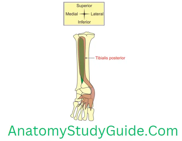
Tibialis posterior muscle
Table of Contents
1. Proximal attachments
1. Upper 2/3rd of lateral part of posterior surface of tibia below the soleal line.
2. Posterior surface of
- Fibula in front of the medial crest, and
- Interosseous membrane.
Read And Learn More: Anatomy Notes And Important Question And Answers
2. Distal attachments
- Tuberosity of navicular bone and other tarsal bones except talus.
- It is extended into 2nd, 3rd and 4th metatarsal bones at their bases.
3. Nerve supply: Tibial nerve segments of spinal cord.
4. Action
- With gastrocnemius and soleus, it brings flexion of foot which acts at ankle joint.
- With tibialis anterior, it brings inversion of foot which acts at subtalar joint, and
- It supports medial longitudinal arch of foot.
Give the attachments and actions of soleus
1. Proximal attachments: It arises from
- The back of head of fibula and upper l/4th of posterior surface of fibula,
- Soleal line and middle l/3rd of medial border of shaft of tibia,
- Deep transverse fascia of leg, and
- Soleal arch which stretches between tibia and fibula.
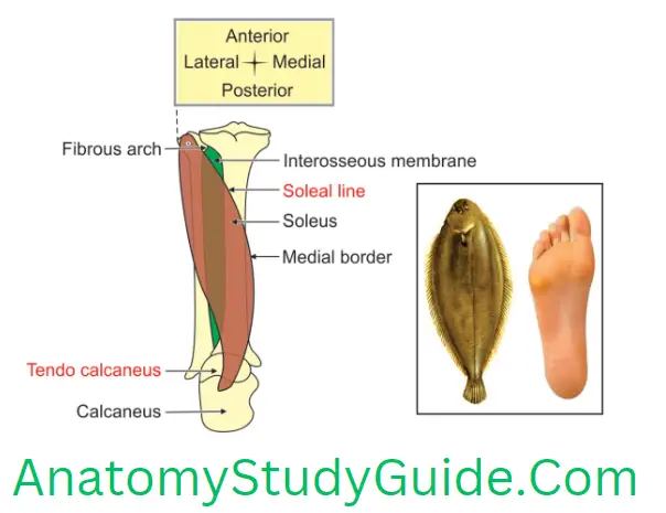
2. Distal attachments: The tendon of soleus fuses with tendon of gastrocnemius to form tendo calcaneus. It is inserted into middle l/3rd of posterior surface of calcaneus.
3. Action
- When the knee is flexed, action of gastrocnemius becomes ineffective. Hence soleus is the main plantar flexor of flexed knee.
- In standing position, it steadies the leg.
- In walking, it acts as a bottom or 1st gear and so it overcomes the inertia of body weight.
Soleus
(Soleus is shaped like the sole of a boot or flat fish (sole fish that reclines on sea floor).)
1. Introduction: It is multipennate superficial muscle of posterior compartment of leg. It is situated deep to the gastrocnemius.
2. Features
- The large size of the gastrosoleus is a human character. It is directly related to the adoption of an erect posture. It is the feature of the bipedal gait of man.
- Morphologically, it corresponds with the flexor digitorum superficialis muscle of the forearm.
- The soleus is located deep to the gastrocnemius and is considered to be the “workhorse” (A horse used for work on a farm) of plantar flexion.
- The gastrocnemius and the soleus are together called the triceps surae which forms the bulging of the calf.
- It forms the 2nd layer of posterior compartment of leg.
- The flexor digitorum brevis is detached distal part of the soleus.
3. Peculiarities
- The soleus contains a rich plexus of small veins. The contraction of the soleus squeezes the vessels and facilitates venous return from the lower extremity. Hence it is often called peripheral heart. The calf muscles collectively work as a venous pump. During relaxed state of the muscles, the blood is sucked from the superficial to the deep veins through the perforators.
- The superficial veins of leg (e.g. saphenous veins) drain into soleal sinuses through perforating veins. The soleal sinuses empty segmentally into deep veins of the leg (e.g. posterior tibial and peroneal veins).
4. Attachments: It arises entirely below knee.
1. Proximal attachment: It has a continuous proximal attachment in the shape of an inverted U. It has two heads.
- Tibial head Soleal line of the tibia, and Middle 1/3rd of the medial border of tibia. Intermuscular septum
- Fibular head: The posterior surfaces of the head of the fibula and of the upper 1/3rd of its shaft;
- These two heads are united by tendinous arch. It represents the upper part of the transverse intermuscular septum and extends between the tibia and the fibula. The additional fibres arise from this arch.
2. Distal attachments: The tendon of soleus fuses with tendon of gastrocnemius and form tendo calcaneus. It is inserted into middle 1/3rd of posterior surface of calcaneus. The tendo calcaneus is thickest and strongest tendon in the body
5. Relations: The popliteal artery and tibial nerve leave the popliteal fossa through proximal attachment of soleus. This bifurcates into its terminal branches, namely the anterior and posterior tibial arteries.
1. Superficial
- Gastrocnemius, and
- Plantaris.
2. Deep
- Flexor digitorum longus,
- Flexor hallucis longus,
- Tibialis posterior,
- Posterior tibial vessels, and
- Tibial nerve.
3. Structures passing deep to soleal arch are popliteal vessels.
6. Blood supply: Sural branch of popliteal artery.
7. Nerve supply : Tibial nerve. The nerves to the soleus, carrying fibres from spinal cord segments, are double.
8. Action
- The soleus and gastrocnemius bring plantar flexion at ankle joint.
- Strong plantar flexion of ankle joint. When the knees flexed, the soleus is the main muscle for plantar flexion at ankle because the action of gastrocnemius becomes ineffective.
- It steadies leg in standing position.
- In walking, it acts as a bottom or 1st gear and so it overcomes the inertia of body weight. It acts as a propelling force in walking.
- Muscle injuries may occur as a result of direct trauma or as part of an overuse syndrome. Tears of the muscles below the knee typically occur within the soleus muscle.
9. Applied anatomy
- Thrombosis or the veins of the soleus muscle give rise to mild pain in the calf. However, deep vein thrombosis (DVT) also occurs with no signs or symptoms. Dislodged thrombus causes pulmonary embolism which is often fatal. Hourly stretching prevents the thrombosis.
- The veins draining the soleus do not have valve. These resemble dural venous sinuses present in the cranium. These are called soleal sinuses.
- The tendo calcaneus is frequently strained at the start of a short race due to abrupt take-off. It may rupture when it is stressed. The complete rupture of this tendon causes abrupt pain in the posterior aspect of the leg and person stumbles with pain. Consequently, he is unable to use his limb and thus cannot walk. He develops lump due to shortening of the gastrocnemius and soleus muscles.
Leave a Reply