Paediatric Periodontics An Overview Introduction
Pediatric periodontics is the science that deals with the upkeep and maintenance of periodontium in primary, mixed, and early permanent dentition. The periodontium comprises the gingiva, periodontal ligament, cementum, and alveolar bone. Th gingiva comprises attached gingiva, marginal gingiva and interdental papilla.
Table of Contents
The periodontium of the primary dentition varies anatomically and microscopically from that of the permanent dentition. The aspects of clinical significance are as follows:
Read And Learn More: Paediatric Dentistry Notes
- Anatomic variations in the periodontium of primary and permanent dentition
- Signifiant gingival features in primary dentition
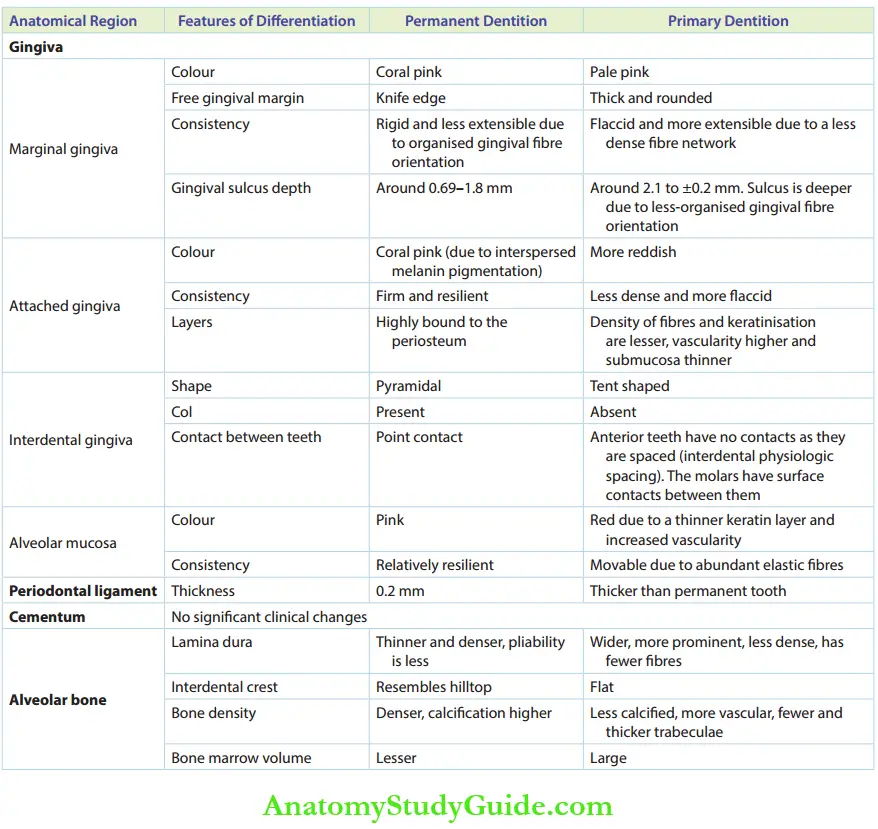
Anatomic Variations Of The Periodontium In Primary And Permanent Dentition
The anatomic variations of the periodontium in primary and permanent dentition are listed in Table.
Significant Gingival Variations In Primary Dentition
There are many features that differentiate the gingiva of primary dentition from those of permanent dentition. The gingiva in primary dentition is more vascular in nature and the gingival fires are less organized when compared to those in permanent dentition. The significant variations are listed and explained as follows:
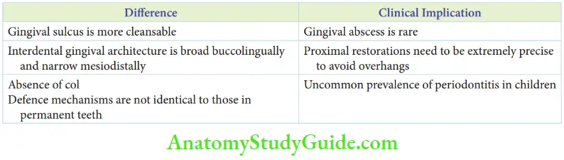
- Interdental gingival architecture: The intern tal gingiva is broad buccolingually and narrower mesiodistally in the primary dentition. This interdental gingival architecture demands precise proximal restorations where minor aberrations in the proximal box contour can lead to overhanging restoration.
- Free gingival margin: Th free gingival margin in primary dentition is thick and rounded. This is in accordance with the cervical bulge and the marked constriction of the primary tooth at the cervix.
- Retractable marginal gingiva: Th marginal gingiva is retractable around the primary tooth. This makes the area more cleansable leaving very less chances for food entrapment. Hence, gingival abscesses are rare in children.
- Interdental cleft: Thy are gingival cleft in the inter-radicular region underlying the saddle area and are more common in the primary dentition.
- Retrocuspid papillae: It is a sessile, elevated, well-circumscribed, sof mucosal nodule, first described by Hirshfeld. It is found in the lingual gingiva of the mandibular cuspids, in between the free gingival margin and the mucogingival junction. It is usually found bilaterally. This structure is exceedingly common in primary dentition with as high as 99% prevalence in the children of age group 8–16. It regresses with age and needs no intervention as it is considered a normal anatomic entity.
- Stippling: It represents the functional adaptation of the gingiva to the masticatory strain indulged in it. Stippling is considered as a sign of healthy gingiva. It is absent in infants and begins to appear at the age of 5 years. It is reported to be found in 35% of children with peak prevalence between 5 and 13 years. Stippling increases in adulthood and progressively declines at old age.
- Absence of col: Col is a para-keratinised or nonkeratinised area beneath the interdental gingiva. It is a tent-shaped region, most susceptible to infection. The col is absent in primary dentition. This is considered as one of the reasons why periodontal diseases are highly uncommon in primary dentition.
Summary
- The differences in periodontium of primary and permanent teeth are listed in Table.
- Anatomic differences between primary and permanent periodontium have significant clinical implications
Leave a Reply