Suprarenal Gland and Chromaffin System
Question – 1: State the two developmental components of the adrenal gland.
Answer:
1. The adrenal cortex develops from mesoderm.
2. The adrenal medulla develops from neural crest cells.
Read And Learn More: General Histology Questions and Answers
Question – 2: What is the structure of the adrenal medulla and what are its secretions?
Answer:
1. Structure of adrenal medulla
Chromaffin cells:
1. They are the most abundant cells in the adrenal medulla.
2. They are ovoid  shaped secretory cells.
3. They are arranged in clumps or cords.
4. They surround the capillaries.
5. The cytoplasm of these cells has secretory granules containing catecholamines (adrenaline and noradrenaline).
6. The secretory granules stain with stains containing chromium salts (chromaffin reaction). Hence, these cells are called chromaffin cells.
7. Chromaffin cells are innervated by preganglionic sympathetic fibers. They correspond functionally to postganglionic sympathetic neurons as both are derived from neural crest cells.
Ganglion cells:
They are singly or in small groups.
They are larger in size than chromaffin cells.
2. Secretions: They produce catecholamines (adrenaline and noradrenaline).
Question 3: What is chromaffin reaction? (Chroma—color, fil—loving)
Answer:
1. The cells of the adrenal medulla have an affinity to color. Hence, these cells are called chromaffin cells.
2. The cytoplasm of these cells has secretory granules containing catecholamines (adrenaline and noradrenaline).
3. The secretory granules are stained with chromium salts. This is called the chromaffin reaction. The cells are called chromaffin cells.
Question 4: Name the hormones secreted by the suprarenal gland.
Answer:
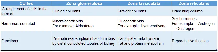
1. Zona glomerulosa: Mineralocorticoids—aldosterone
2. Zona fasciculate: Hydrocorticoids—hydrocortisone
3. Zona reticularis: Sex hormone—androgen and estrogen
Question – 5: Describe the suprarenal gland under the following heads
1. Gross anatomy
2. Histology
3. Development, and
4. Applied anatomy.
Answer:
Introduction:
Shape:
- The right suprarenal gland is
 lar or irregular tetrahedron.
 - Left suprarenal gland is
 semilunar.
Dimensions:
- Right suprarenal gland is 4 x 4 x 1 cm
- Left suprarenal gland is 5 x 3 x 1 cm
Weight: 5 g
Situation:
- The right suprarenal gland is situated at the upper pole of the right kidney.
- It covers the anterior surface of the right kidney.
- The left suprarenal gland is situated on the upper part of the medial border of the left kidney.
1. Gross anatomy
1. External features:
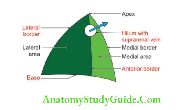
The right suprarenal gland has:
1. Apex
2. Base
3. Two surfaces
- Anterior and
- Posterior, and
4. Two borders
- Medial, and
- Lateral.
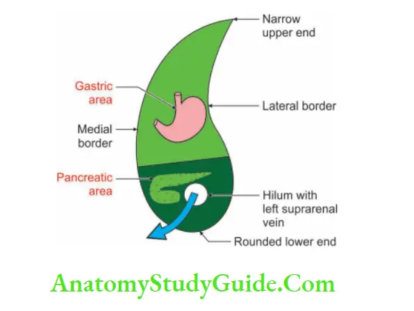
The left suprarenal gland has:
1. Two ends
- Upper and
- Lower
2. Two borders
- Medial and
- Lateral, and
3. Two surfaces
- Anterior and
- Posterior.
Hilum of the suprarenal gland:
1. Right hilum:
Situation: Anterior surface of the upper pole of the right kidney
Structures leaving:
- Right suprarenal vein, and
- Lymph vessels.
2. Left hilum:
Situation: Anterior surface and near the lower pole of the left suprarenal gland.
Structures leaving:
- Left suprarenal vein, and
- Lymph vessels
Hat Ke:
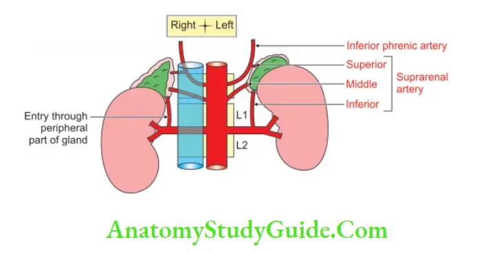
Note: Suprarenal arteries do not enter through the hilum of the suprarenal gland.
2. Relations:
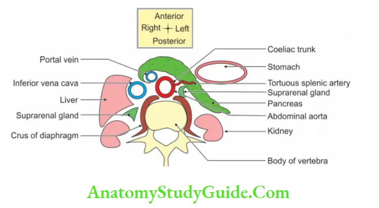
Right suprarenal gland:
1. Anterior surface: It has medial and lateral parts
Medial part: Inferior vena cava (no peritoneum).
Lateral part: It is related to the liver and is not covered by the peritoneum.
- The bare area of the liver (no peritoneum).
- Inferior surface of the liver (peritoneal).
2. Posterior surface: Divided into lateral and medial parts by crest.
Lateral part: Anterior surface of the right kidney.
Medial part: Right crus of the diaphragm.
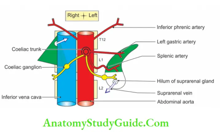
3. Medial border:
- Right inferior phrenic artery, and
- Right coeliac ganglion.
Left suprarenal gland:
1. Anterior surface:
- The upper part is covered with the peritoneum and related to the posterior surface of the stomach.
- The lower part is non-peritoneal overlapped by the body of the pancreas and crossed by the tortuous splenic artery.
2. Posterior surface:
- Left kidney, and
- Left crus of the diaphragm.
3. Medial border:
- Left coeliac ganglion
- Left inferior phrenic artery, and
- Left gastric artery.
- Lateral border: Left kidney.
3. Blood supply:
Arterial supply: It receives blood from the following arteries. All arteries enter through the peripheral part of the gland, i.e. cortex, not through the hilum.
- Superior suprarenal artery, a branch of the inferior phrenic artery.
- Middle suprarenal artery, a branch of the abdominal aorta.
- Inferior suprarenal artery, is a branch of the renal artery.
Venous drainage:
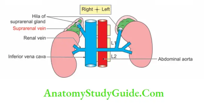
1. The veins of the suprarenal gland drain in the following veins:
- The right suprarenal vein drains the inferior vena cava.
- The left suprarenal vein drains into the left renal vein.
2. Peculiarities of the veins draining the suprarenal gland:
- There is only one thick vein equal to the diameter of the lead pencil.
- The reason to have only one vein is to have the uniform distribution of the hormone to all parts of the gland.
- The vein draining the suprarenal gland leaves through the hilum.
- The left suprarenal vein is longer as compared to the right suprarenal vein.
5. Nerve supply:
1. Cortex: The nerve supply of the cortex is not known.
2. Medulla is supplied by
- Sympathetic (thoracic splanchnic nerve). The pre-ganglionic fibres, directly go to the cells of the medulla.
- Phrenic, and
- Vagus nerves
Know what is not known:
Note: The exact functions of phrenic and vagus nerves are not known.
6. Lymphatic drainage: The lymphatics of the suprarenal glands may accompany any vessel reaching the adrenal gland. The lymphatics drain into the paraaortic nodes.
- The right para-aortic nodes are situated near the right crus of the diaphragm.
- The left para-aortic nodes are situated near the origin of the left renal artery.
2. Histology
Divided into cortex and medulla.
1. Cortex: The cortex is divided into three zones
Zones of the cortex of the suprarenal gland:
2. Adrenal medulla: Cells of the medulla exhibit chromaffin reaction, i.e. when treated with potassium dichromate solution, the cytoplasmic granules are oxidized. The medulla turns brown.
It shows
1. Cords with pheochromocytes (pheo—dusky or brown, chroma—colors, cytes— cell). They are of two types.
- Adrenaline secreting.
- Noradrenaline secreting.
2. A few sympathetic ganglion cells show prominent vesicular nucleoli.
3. Sinusoidal capillaries present between cell groups.
3. Development
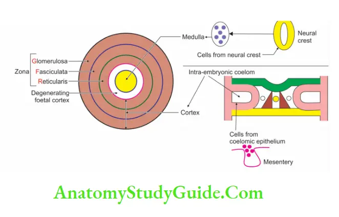
Chronological age: Adrenal cortex develops in the 6th week of intrauterine life.
Germ layer: Inside out
- The cortex develops from the mesoderm.
- The medulla develops from the ectoderm.
Site: Between dorsal mesentery and developing gonad.
Sources: In the fetus, the suprarenal is 20 times larger than the adult because of the large fetal cortex which regresses after birth.
- Cortex develops from coelomic epithelium.
- The medulla develops from neural crest (neuroectoderm).
Anomalies:
1. Abnormal sites:
- Deep to the capsule of the kidney.
- It may be fused to the liver or kidney.
2. Congenital adrenal hyperplasia: An abnormal increase in the cells of the suprarenal cortex.
In males ♂: it leads to adrenogenital syndrome. It is made by the early development of secondary sexual characters.
Female ♀:
- It causes enlargement of the clitoris. The child may be mistaken for pseudo-hermaphroditism.
- It produces excessive androgen during the fetal period. It results in the masculinization of females.
4. Applied anatomy
Addison’s disease: It is caused by atrophy of the suprarenal cortex. It is mainly due to tuberculosis infection. It presents as
- Muscular weakness
- Low blood pressure
- Cutaneous pigmentation, and
- Change in electrolyte balance.
2. Pheochromocytoma (the—grey, chroma—color, cyt—cell, oma—tumor):
It is a tumor of the adrenal medulla, that produces paroxysmal hypertension due to the secretion of large amounts of catecholamine. It produces
- Palpitation
- Excessive sweating
- Pallor
- Hypertension, and
- Headache of long duration
- The adrenal medulla can be sacrificed, and the secretions are replaced by medicines.

Leave a Reply