Tooth Preparation To Receive All Ceramic Crown Introduction
All ceramic crowns are considered as esthetic option for anterior restoration. The preparation to receive all ceramic crowns needs more amount of tooth reduction compared to other available options. This chapter describes stepwise tooth preparation to receive all ceramic crown restoration.
Table of Contents
Tooth Preparation
The stepwise preparation of maxillary central incisor to receive all ceramic crowns is given here.
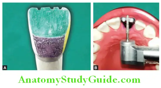
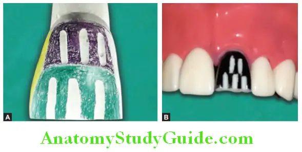
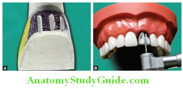
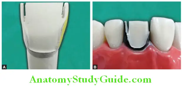
Incisal Reduction:
Depth of preparation (amount of tooth preparation): 2 mm.
Instrument: Flat-end tapering diamond. Place depths orienting grooves or depth cuts on the incisal edge. Place three cuts on incisal edge, one on mid-incisal region and one each at the junction of each proximal surface. Remove remaining tooth structure from incisal edge using small wheel diamond or flat-end tapering diamond.
Read and Learn More: Preclinical Prosthodontics Notes
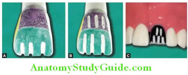
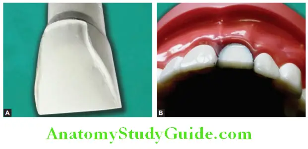
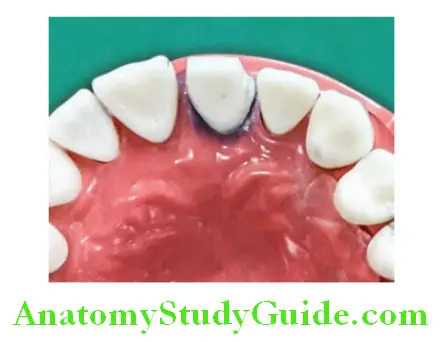
Labial Reduction:
Depth of preparation (amount of tooth preparation): 1.5 mm.
Instrument: Flat-end tapering diamond. The labial surface of incisor can be divided in two planes. These are located at different angulation thus labial surface is prepared in two planes.
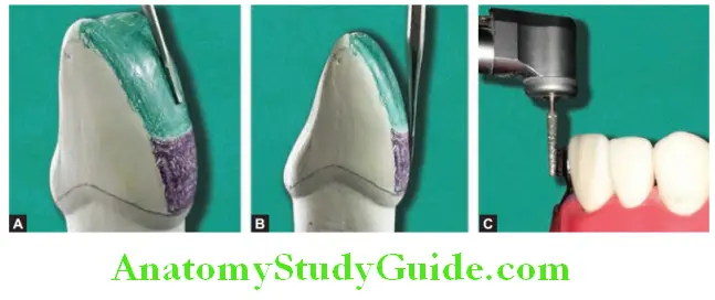
Prepare the labial surface of maxillary incisor in two planes. Place the depth orientation grooves by sinking flat-end tapering diamond up to 1.2 mm which after finishing will produce a depth of 1.5 mm. Place the depth orienting grooves in two planes.
One set within the gingival half and it is parallel to gingival half of labial surface. The other set is placed in incisal half. Remove the remaining tooth structure between the depth grooves using the flat-end tapering diamond. Extend the preparation to the facioproximal line angle.
Follow gingival contour during tooth preparation. This will prevent trauma to interdental papillae and limit the preparation in gingival sulcus. A shoulder finish line is produced by flatend tapered diamond. Shoulder finish line can be placed subgingival (recommended for anterior teeth) or equigingival.
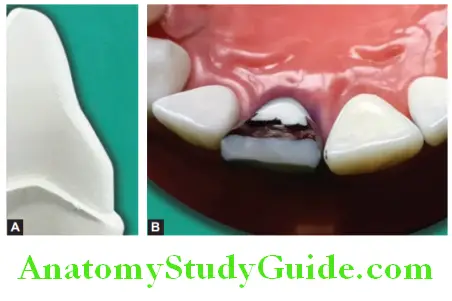
Lingual Reduction:
Lingual reduction is done in two parts. Axial reduction is done first and then tooth reduction done from lingual fossa.
Lingual Axial Reduction:
Depth of preparation (amount of tooth preparation): 1.5 mm.
Instrument:
Flat-end tapering diamond. Lingual axial reduction will prepare cervical portion of lingual surface of tooth. This surface should be given taper of 3–5° with cervical part of labial surface. Place flat end taper diamond straight against this surface and prepare the tooth. Due to taper configuration of bur, the surface will be prepared in tapered style. Maintain the position of bur and extend tooth preparation till linguoproximal line angle. A shoulder finish line is recommended in lingual surface.
Lingual Fossa Reduction:
Depth of preparation (amount of tooth preparation): 1.5 mm.
Instrument: Number 6 round bur and wheel diamond or football diamond. Place depth cut in form of pot holes in lingual fossa with a number 6 round bur. The diameter of round bur should be 1.8 mm which produces depth cuts of approximately 1–1.2 mm after finishing it gives required depth of preparation. The remaining tooth structure is removed with small wheel diamond or football diamond.
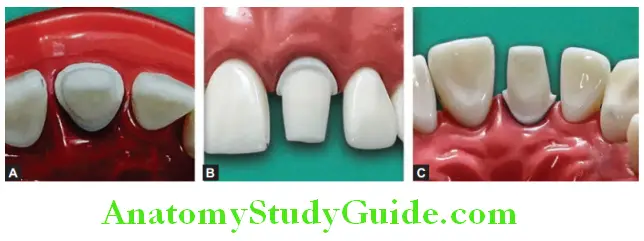
Proximal Reduction:
Depth of preparation (amount of tooth preparation): 1.5 mm.
Instrument: Long thin tapering diamond and flat-end tapering diamond. Long thin tapering diamond is used to prepare proximal tooth surface. The access is prepared using a vertical sawing motion. The diamond should follow incisogingival direction.
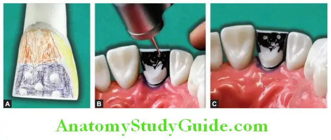
This motion will cut the tooth surface proximally. The cut is through proximal enamel and produce the enamel lip.
Remove enamel lip by any hand instrument or by diamond point. Use flat-end tapering diamond to join facial and lingual preparation through proximal area. This produces proximal surface with taper of 3–5°. Produce smooth shoulder in proximal area following the gingival contour. Prepared tooth to receive all ceramic crown.
Finishing Of Preparation
Make all the point and line angles rounded to prevent stress concentration and fracture. Axial surface can be finished with yellow collar fine grit diamond. The shoulder can
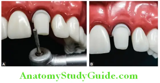
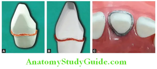
be finished with end cutting diamond where only tip has fine diamonds and sides are noncutting. See the shoulder finish line on labial side, proximal side, and lingual side.
Leave a Reply