Tooth Preparation To Receive All Metal Crown Introduction
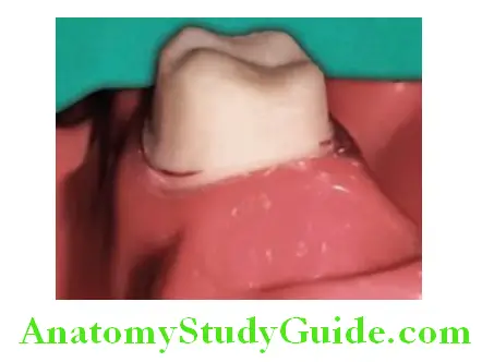
The preparation to receive all metal crown needs less amount of tooth reduction. This chapter describes stepwise tooth preparation to receive all metal crown restoration on mandibular molar.
Table of Contents
Tooth Preparation
The stepwise preparation of maxillary central incisor to receive porcelain fused metal (PFM) crown is given here.
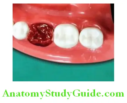
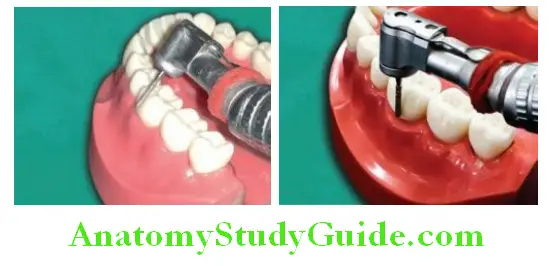
Occlusal Reduction:
Depth of preparation (amount of tooth preparation): 1 mm on nonfunctional cusp and 1.5 mm on functional cusp.
Instrument: Round-end tapering diamond. Depth orienting grooves or depth cuts of 1 mm are placed on the occlusal grooves. Round diamond or bur of 2 mm diameter can also be used for placing depth cuts on occlusal surface. Place depth cuts on triangular ridges from cusp tip to base. Remove tooth structure between the depth cuts. Follow the contour and anatomy during tooth preparation.
Read and Learn More: Preclinical Prosthodontics Notes
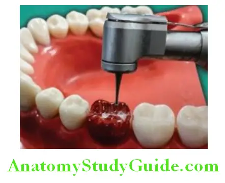
Axial Reduction: Buccal and Lingual Preparation:
Depth of preparation (amount of tooth preparation): 0.8–1 mm on buccal surface. On cervical one-third, the preparation depth is 0.5 mm.
Instrument: Round-end tapering diamond. The buccal surface of mandibular molar is prepared in two planes. The depth orienting grooves are placed by sinking round-end tapering diamond up to half thickness (if the diameter of bur is 2 mm, than it will place depth orientation grooves of 1 mm
by sinking the bur by half thickness). The depth orienting grooves are placed in two planes. One within the gingival half and it is parallel to gingival half of axial surface. The other is placed in occlusal half. The remaining tooth structure between the depth grooves is then removed using the round-end tapering diamond. Extend the preparation proximally 1 mm beyond the contact point Follow gingival contour during tooth preparation. This will prevent trauma to interdental papillae.
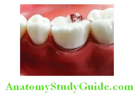
Preparing the buccal surface with roundend tapering diamond produces necessary taper of 3–5°.
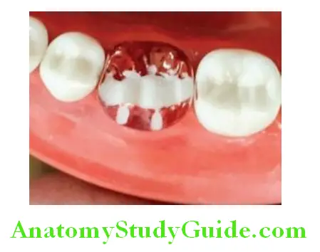
Proximal Reduction:
Depth of preparation (amount of tooth preparation): 0.8–1 mm.
Instrument: Long thin tapering diamond and round-end tapering diamond. Long thin tapering diamond is used to prepare proximal tooth surface. The access is prepared using a vertical sawing motion.
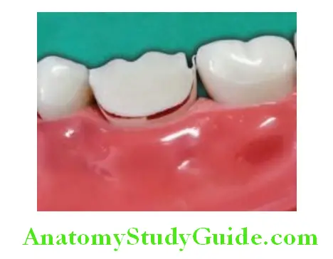
The diamond should follow incisogingival direction. This motion will cut the tooth surface proximally. The cut is through proximal enamel and produce the enamel lip. Remove enamel lip by any hand instrument or by diamond point.
Use round-end tapering diamond to join facial and lingual preparation through proximal area. This produces proximal surface with taper of 3–5°. Produce smooth chamfer in proximal area following the gingival contour.
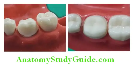
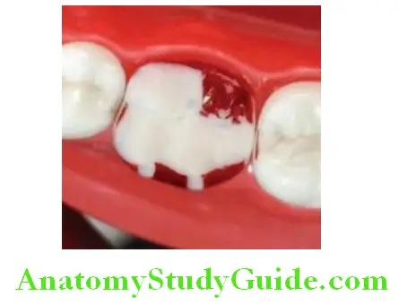
Functional Cusp Bevel:
Functional cusp bevel is prepared at 45° to axial surface. This is prepared to give extra bulk of material on functional cusp to prevent fracture of restoration and to increase structural durability. Functional cusp bevel is given by round-end tapering diamond placing at 45° to functional cusps.
Finishing Of Preparation
Make all the point and line angles rounded. Axial surface can be finished with torpedo fine grit diamond.
Leave a Reply